 Movie
Movie Controller


+ Open data

- Basic information
| Entry | Database: PDB / ID: 6uga | ||||||
|---|---|---|---|---|---|---|---|
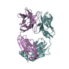
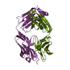
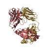
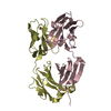
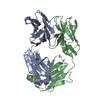
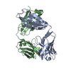
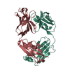
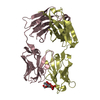
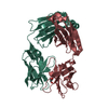
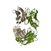
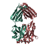
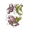
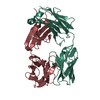
| Title | ch28/11 Fab (monoclinic form) | ||||||
 Components Components |
| ||||||
 Keywords Keywords | IMMUNE SYSTEM / IMMUNOGLOBULIN / CHIMERIC ANTIBODY / ANTIGEN BINDING FRAGMENT | ||||||
| Function / homology | Immunoglobulins / Immunoglobulin-like / Sandwich / Mainly Beta / methanesulfonic acid Function and homology information Function and homology information | ||||||
| Biological species |  | ||||||
| Method |  X-RAY DIFFRACTION / SYNCHROTRON / MOLECULAR REPLACEMENT / Resolution: 2.5 Å X-RAY DIFFRACTION / SYNCHROTRON / MOLECULAR REPLACEMENT / Resolution: 2.5 Å | ||||||
 Authors Authors | Soliman, C. / Ramsland, P.A. | ||||||
 Citation Citation | Journal: J.Biol.Chem. / Year: 2020 Title: The terminal sialic acid of stage-specific embryonic antigen-4 has a crucial role in binding to a cancer-targeting antibody. Authors: Soliman, C. / Chua, J.X. / Vankemmelbeke, M. / McIntosh, R.S. / Guy, A.J. / Spendlove, I. / Durrant, L.G. / Ramsland, P.A. | ||||||
| History |
|
- Structure visualization
| Structure viewer | Molecule: MolmilJmol/JSmol |
|---|
- Downloads & links
-Download
| PDBx/mmCIF format | 6uga.cif.gz | 173.8 KB | Display | PDBx/mmCIF format |
|---|---|---|---|---|
| PDB format | pdb6uga.ent.gz | 136.9 KB | Display | PDB format |
| PDBx/mmJSON format | 6uga.json.gz | Tree view | PDBx/mmJSON format | |
| Others |  Other downloads Other downloads |
-Validation report
| Arichive directory | https://data.pdbj.org/pub/pdb/validation_reports/ug/6ugaftp://data.pdbj.org/pub/pdb/validation_reports/ug/6uga | HTTPS FTP |
|---|
-Related structure data
| Related structure data | 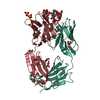 6ug7SC 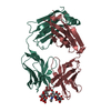 6ug8C 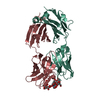 6ug9C S: Starting model for refinement C: citing same article ( |
|---|---|
| Similar structure data |


-Links
 PDBj
PDBj

- Assembly
| Deposited unit | 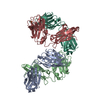
| ||||||||
|---|---|---|---|---|---|---|---|---|---|
| 1 | 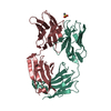
| ||||||||
| 2 | 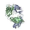
| ||||||||
| Unit cell |
|
-Components
| #1: Antibody | Mass: 23166.750 Da / Num. of mol.: 2 Source method: isolated from a genetically manipulated source Source: (gene. exp.)  Homo sapiens (human) Homo sapiens (human)#2: Antibody | Mass: 23016.844 Da / Num. of mol.: 2 Source method: isolated from a genetically manipulated source Source: (gene. exp.) Homo sapiens (human)#3: Chemical | ChemComp-03S / |   Mass: 96.106 Da / Num. of mol.: 1 / Source method: obtained synthetically / Formula: CH4O3S Mass: 96.106 Da / Num. of mol.: 1 / Source method: obtained synthetically / Formula: CH4O3S#4: Water | ChemComp-HOH / |  Mass: 18.015 Da / Num. of mol.: 119 / Source method: isolated from a natural source / Formula: H2O Mass: 18.015 Da / Num. of mol.: 119 / Source method: isolated from a natural source / Formula: H2OHas ligand of interest | N | Has protein modification | Y | |
|---|
-Experimental details
-Experiment
| Experiment | Method: X-RAY DIFFRACTION / Number of used crystals: 1 |
|---|
- Sample preparation
| Crystal | Density Matthews: 2.7 Å3/Da / Density % sol: 53.9 % |
|---|---|
| Crystal grow | Temperature: 291 K / Method: vapor diffusion, hanging drop / pH: 7.5 / Details: PEG 4000, iso-Propanol, sodium HEPES |
-Data collection
| Diffraction | Mean temperature: 100 K / Serial crystal experiment: N |
|---|---|
| Diffraction source | Source: SYNCHROTRON / Site: Australian Synchrotron  / Beamline: MX2 / Wavelength: 0.954 Å / Beamline: MX2 / Wavelength: 0.954 Å |
| Detector | Type: DECTRIS EIGER X 16M / Detector: PIXEL / Date: Jun 29, 2019 |
| Radiation | Protocol: SINGLE WAVELENGTH / Monochromatic (M) / Laue (L): M / Scattering type: x-ray |
| Radiation wavelength | Wavelength: 0.954 Å / Relative weight: 1 |
| Reflection | Resolution: 2.5→50 Å / Num. obs: 33478 / % possible obs: 98.1 % / Redundancy: 3.4 % / Rrim(I) all: 0.07 / Net I/σ(I): 14.4 |
| Reflection shell | Resolution: 2.5→2.6 Å / Num. unique obs: 5126 / Rrim(I) all: 0.48 |
- Processing
| Software |
| |||||||||||||||||||||||||||||||||||||||||||||||||||||||||||||||||||||||||||
|---|---|---|---|---|---|---|---|---|---|---|---|---|---|---|---|---|---|---|---|---|---|---|---|---|---|---|---|---|---|---|---|---|---|---|---|---|---|---|---|---|---|---|---|---|---|---|---|---|---|---|---|---|---|---|---|---|---|---|---|---|---|---|---|---|---|---|---|---|---|---|---|---|---|---|---|---|
| Refinement | Method to determine structure: MOLECULAR REPLACEMENT Starting model: 6UG7 Resolution: 2.5→50 Å / Cor.coef. Fo:Fc: 0.966 / Cor.coef. Fo:Fc free: 0.929 / Cross valid method: THROUGHOUT / σ(F): 0 / ESU R: 0.473 / ESU R Free: 0.279 Details: HYDROGENS HAVE BEEN ADDED IN THE RIDING POSITIONS U VALUES : REFINED INDIVIDUALLY
| |||||||||||||||||||||||||||||||||||||||||||||||||||||||||||||||||||||||||||
| Solvent computation | Ion probe radii: 0.8 Å / Shrinkage radii: 0.8 Å / VDW probe radii: 1.2 Å | |||||||||||||||||||||||||||||||||||||||||||||||||||||||||||||||||||||||||||
| Displacement parameters | Biso max: 142.24 Å2 / Biso mean: 58.7148 Å2 / Biso min: 29.24 Å2
| |||||||||||||||||||||||||||||||||||||||||||||||||||||||||||||||||||||||||||
| Refinement step | Cycle: LAST / Resolution: 2.5→50 Å
| |||||||||||||||||||||||||||||||||||||||||||||||||||||||||||||||||||||||||||
| Refine LS restraints |
| |||||||||||||||||||||||||||||||||||||||||||||||||||||||||||||||||||||||||||
| LS refinement shell | Resolution: 2.5→2.565 Å / Total num. of bins used: 20
|
