 Movie
Movie Controller

[English] 日本語
 Yorodumi
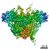
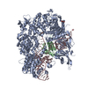
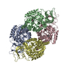
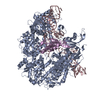
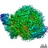
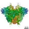
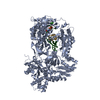
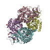
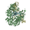
- PDB-6uen: Cryo-EM structure of the respiratory syncytial virus RNA polymerase -
+ Open data

- Basic information
| Entry | Database: PDB / ID: 6uen | ||||||
|---|---|---|---|---|---|---|---|
| Title | Cryo-EM structure of the respiratory syncytial virus RNA polymerase | ||||||
 Components Components |
| ||||||
 Keywords Keywords | VIRAL PROTEIN/Transferase / Viral protein complexes / VIRAL PROTEIN / VIRAL PROTEIN-Transferase complex | ||||||
| Function / homology |  Function and homology information Function and homology informationRespiratory syncytial virus genome transcription / Translation of respiratory syncytial virus mRNAs / GDP polyribonucleotidyltransferase / negative stranded viral RNA replication / Respiratory syncytial virus genome replication / RSV-host interactions / Assembly and release of respiratory syncytial virus (RSV) virions / Maturation of hRSV A proteins / Respiratory syncytial virus (RSV) attachment and entry / Hydrolases; Acting on acid anhydrides; In phosphorus-containing anhydrides ...Respiratory syncytial virus genome transcription / Translation of respiratory syncytial virus mRNAs / GDP polyribonucleotidyltransferase / negative stranded viral RNA replication / Respiratory syncytial virus genome replication / RSV-host interactions / Assembly and release of respiratory syncytial virus (RSV) virions / Maturation of hRSV A proteins / Respiratory syncytial virus (RSV) attachment and entry / Hydrolases; Acting on acid anhydrides; In phosphorus-containing anhydrides / viral life cycle / virion component / symbiont-mediated suppression of host NF-kappaB cascade / host cell cytoplasm / mRNA 5'-cap (guanine-N7-)-methyltransferase activity / RNA-directed RNA polymerase / hydrolase activity / RNA-directed RNA polymerase activity / GTPase activity / ATP binding / metal ion binding Similarity search - Function | ||||||
| Biological species |  Human respiratory syncytial virus Human respiratory syncytial virus | ||||||
| Method | ELECTRON MICROSCOPY / single particle reconstruction / cryo EM / Resolution: 3.67 Å | ||||||
 Authors Authors | Cao, D. / Gao, Y. / Liang, B. | ||||||
| Funding support |  United States, 1items United States, 1items
| ||||||
 Citation Citation | Journal: Nat Commun / Year: 2020 Title: Cryo-EM structure of the respiratory syncytial virus RNA polymerase. Authors: Dongdong Cao / Yunrong Gao / Claire Roesler / Samantha Rice / Paul D'Cunha / Lisa Zhuang / Julia Slack / Mason Domke / Anna Antonova / Sarah Romanelli / Shayon Keating / Gabriela Forero / ...Authors: Dongdong Cao / Yunrong Gao / Claire Roesler / Samantha Rice / Paul D'Cunha / Lisa Zhuang / Julia Slack / Mason Domke / Anna Antonova / Sarah Romanelli / Shayon Keating / Gabriela Forero / Puneet Juneja / Bo Liang / Abstract: The respiratory syncytial virus (RSV) RNA polymerase, constituted of a 250 kDa large (L) protein and tetrameric phosphoprotein (P), catalyzes three distinct enzymatic activities - nucleotide ...The respiratory syncytial virus (RSV) RNA polymerase, constituted of a 250 kDa large (L) protein and tetrameric phosphoprotein (P), catalyzes three distinct enzymatic activities - nucleotide polymerization, cap addition, and cap methylation. How RSV L and P coordinate these activities is poorly understood. Here, we present a 3.67 Å cryo-EM structure of the RSV polymerase (L:P) complex. The structure reveals that the RNA dependent RNA polymerase (RdRp) and capping (Cap) domains of L interact with the oligomerization domain (P) and C-terminal domain (P) of a tetramer of P. The density of the methyltransferase (MT) domain of L and the N-terminal domain of P (P) is missing. Further analysis and comparison with other RNA polymerases at different stages suggest the structure we obtained is likely to be at an elongation-compatible stage. Together, these data provide enriched insights into the interrelationship, the inhibitors, and the evolutionary implications of the RSV polymerase. | ||||||
| History |
|
- Structure visualization
| Movie |
Movie viewer |
|---|---|
| Structure viewer | Molecule: MolmilJmol/JSmol |
- Downloads & links
-Download
| PDBx/mmCIF format | 6uen.cif.gz | 319.4 KB | Display | PDBx/mmCIF format |
|---|---|---|---|---|
| PDB format | pdb6uen.ent.gz | 250.4 KB | Display | PDB format |
| PDBx/mmJSON format | 6uen.json.gz | Tree view | PDBx/mmJSON format | |
| Others |  Other downloads Other downloads |
-Validation report
| Arichive directory | https://data.pdbj.org/pub/pdb/validation_reports/ue/6uenftp://data.pdbj.org/pub/pdb/validation_reports/ue/6uen | HTTPS FTP |
|---|
-Related structure data
| Related structure data | 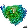 20754MC M: map data used to model this data C: citing same article ( |
|---|---|
| Similar structure data |

-Links
 PDBj
PDBj
- Assembly
| Deposited unit | 
|
|---|---|
| 1 |
|
-Components
| #1: Protein | Mass: 173586.594 Da / Num. of mol.: 1 Source method: isolated from a genetically manipulated source Source: (gene. exp.) Human respiratory syncytial virus / Gene: L, MZ07_64039gpL, MZ07_64040gpL / Plasmid: pET / Cell line (production host): Sf21 / Production host:   Spodoptera frugiperda (fall armyworm) Spodoptera frugiperda (fall armyworm)References: UniProt: G8EJ12, UniProt: P28887*PLUS, RNA-directed RNA polymerase, mRNA (guanine-N7)-methyltransferase, Transferases; Transferring phosphorus-containing groups; Nucleotidyltransferases, ...References: UniProt: G8EJ12, UniProt: P28887*PLUS, RNA-directed RNA polymerase, mRNA (guanine-N7)-methyltransferase, Transferases; Transferring phosphorus-containing groups; Nucleotidyltransferases, GDP polyribonucleotidyltransferase |
|---|---|
| #2: Protein | Mass: 27165.838 Da / Num. of mol.: 4 Source method: isolated from a genetically manipulated source Source: (gene. exp.) Human respiratory syncytial virus / Cell line (production host): Sf21 / Production host: Spodoptera frugiperda (fall armyworm) / References: UniProt: G3C7Q7, UniProt: P03421*PLUS |
-Experimental details
-Experiment
| Experiment | Method: ELECTRON MICROSCOPY |
|---|---|
| EM experiment | Aggregation state: PARTICLE / 3D reconstruction method: single particle reconstruction |
- Sample preparation
| Component | Name: the L protein of the human respiratory syncytial virus; the phosphoprotein (P) of human respiratory syncytial virus Type: COMPLEX / Entity ID: all / Source: RECOMBINANT |
|---|---|
| Molecular weight | Value: 0.360 MDa / Experimental value: YES |
| Source (natural) | Organism: Human respiratory syncytial virus |
| Source (recombinant) | Organism: Spodoptera frugiperda (fall armyworm) / Strain: Sf21 |
| Buffer solution | pH: 7.4 |
| Specimen | Embedding applied: NO / Shadowing applied: NO / Staining applied: NO / Vitrification applied: YES |
| Vitrification | Cryogen name: ETHANE / Humidity: 91 % |
- Electron microscopy imaging
| Experimental equipment |  Model: Talos Arctica / Image courtesy: FEI Company |
|---|---|
| Microscopy | Model: FEI TALOS ARCTICA |
| Electron gun | Electron source:  FIELD EMISSION GUN / Accelerating voltage: 200 kV / Illumination mode: FLOOD BEAM FIELD EMISSION GUN / Accelerating voltage: 200 kV / Illumination mode: FLOOD BEAM |
| Electron lens | Mode: BRIGHT FIELD |
| Image recording | Electron dose: 55 e/Å2 / Film or detector model: GATAN K2 SUMMIT (4k x 4k) |
- Processing
| Software | Name: PHENIX / Version: 1.11.1_2575: / Classification: refinement | ||||||||||||||||||||||||
|---|---|---|---|---|---|---|---|---|---|---|---|---|---|---|---|---|---|---|---|---|---|---|---|---|---|
| CTF correction | Type: PHASE FLIPPING AND AMPLITUDE CORRECTION | ||||||||||||||||||||||||
| 3D reconstruction | Resolution: 3.67 Å / Resolution method: FSC 0.143 CUT-OFF / Num. of particles: 253372 / Symmetry type: POINT | ||||||||||||||||||||||||
| Atomic model building | Protocol: AB INITIO MODEL / Space: REAL | ||||||||||||||||||||||||
| Refine LS restraints |
|
