[English] 日本語
Yorodumi
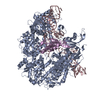
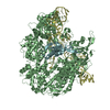
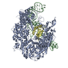
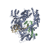
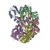
- PDB-5vw1: Crystal structure of SpyCas9-sgRNA-AcrIIA4 ternary complex -

+
Open data


ID or keywords:

Loading...

-
Basic information

Entry
Database: PDB / ID: 5vw1
TitleCrystal structure of SpyCas9-sgRNA-AcrIIA4 ternary complex
Components
  • CRISPR-associated endonuclease Cas9/Csn1
  • anti-CRISPR protein AcrIIA4
  • sgRNASubgenomic mRNA
KeywordsHYDROLASE / Type II CRISPR-Cas endonculease: Cas9: Structure: Endonuclease: anti-CRISPR protein: Inhibition of Cas9: RuvC catalytic pocket: Sequence-specific PAM recognition: Genome editing tool
Function / homology
Function and homology information


maintenance of CRISPR repeat elements / 3'-5' exonuclease activity / DNA endonuclease activity / defense response to virus / Hydrolases; Acting on ester bonds / DNA binding / RNA binding / metal ion binding
Similarity search - Function
CRISPR-associated endonuclease Cas9, PAM-interacting domain / CRISPR-associated endonuclease Cas9, bridge helix / CRISPR-associated endonuclease Cas9, REC lobe / REC lobe of CRISPR-associated endonuclease Cas9 / Bridge helix of CRISPR-associated endonuclease Cas9 / PAM-interacting domain of CRISPR-associated endonuclease Cas9 / CRISPR-associated endonuclease Cas9 / HNH endonuclease / Cas9-type HNH domain / Cas9-type HNH domain profile. ...CRISPR-associated endonuclease Cas9, PAM-interacting domain / CRISPR-associated endonuclease Cas9, bridge helix / CRISPR-associated endonuclease Cas9, REC lobe / REC lobe of CRISPR-associated endonuclease Cas9 / Bridge helix of CRISPR-associated endonuclease Cas9 / PAM-interacting domain of CRISPR-associated endonuclease Cas9 / CRISPR-associated endonuclease Cas9 / HNH endonuclease / Cas9-type HNH domain / Cas9-type HNH domain profile. / HNH nuclease / Ribonuclease H superfamily
Similarity search - Domain/homology
DI(HYDROXYETHYL)ETHER / RNA / RNA (> 10) / Uncharacterized protein / CRISPR-associated endonuclease Cas9/Csn1
Similarity search - Component
Biological speciesStreptococcus pyogenes serotype M1 (bacteria)
Listeria monocytogenes serotype 4a
Listeria monocytogenes (bacteria)
MethodX-RAY DIFFRACTION / SYNCHROTRON / MOLECULAR REPLACEMENT / Resolution: 2.598 Å
AuthorsYang, H. / Patel, D.J.
Funding support United States, 2items
OrganizationGrant numberCountry
National Institutes of Health/National Institute of General Medical Sciences (NIH/NIGMS)GM104962 United States
Memorial Sloan Kettering Cancer Center Core GrantP30CA008748 United States
CitationJournal: Mol. Cell / Year: 2017
Title: Inhibition Mechanism of an Anti-CRISPR Suppressor AcrIIA4 Targeting SpyCas9.
Authors: Yang, H. / Patel, D.J.
History
DepositionMay 21, 2017Deposition site: RCSB / Processing site: RCSB
Revision 1.0Jun 28, 2017Provider: repository / Type: Initial release
Revision 1.1Jul 19, 2017Group: Database references / Category: citation
Item: _citation.journal_volume / _citation.page_first / _citation.page_last
Revision 1.2Sep 20, 2017Group: Author supporting evidence / Category: pdbx_audit_support / Item: _pdbx_audit_support.funding_organization
Revision 1.3Jan 1, 2020Group: Author supporting evidence / Category: pdbx_audit_support / Item: _pdbx_audit_support.funding_organization
Revision 1.4Oct 4, 2023Group: Data collection / Database references / Refinement description
Category: chem_comp_atom / chem_comp_bond ...chem_comp_atom / chem_comp_bond / database_2 / pdbx_initial_refinement_model
Item: _database_2.pdbx_DOI / _database_2.pdbx_database_accession

-
Structure visualization

Structure viewerMolecule:
MolmilJmol/JSmol

Downloads & links

-
Assembly

Deposited unit
A: CRISPR-associated endonuclease Cas9/Csn1
C: sgRNA
B: anti-CRISPR protein AcrIIA4
hetero molecules


Theoretical massNumber of molelcules
Total (without water)197,1469
Polymers196,4593
Non-polymers6876
Water3,423190
1


  • Idetical with deposited unit
  • defined by author&software
  • Evidence: gel filtration
TypeNameSymmetry operationNumber
identity operation1_555x,y,z1
Buried area16090 Å2
ΔGint-88 kcal/mol
Surface area73910 Å2
MethodPISA
Unit cell
Length a, b, c (Å)72.194, 101.242, 303.563
Angle α, β, γ (deg.)90.00, 90.00, 90.00
Int Tables number18
Space group name H-MP22121

-
Components

-
Protein , 2 types, 2 molecules AB

#1: Protein CRISPR-associated endonuclease Cas9/Csn1 / SpCas9 / SpyCas9


Mass: 158732.891 Da / Num. of mol.: 1 / Mutation: D10A/H840A
Source method: isolated from a genetically manipulated source
Source: (gene. exp.) Streptococcus pyogenes serotype M1 (bacteria)
Gene: cas9, csn1, SPy_1046 / Plasmid: pRSFDuet-SUMO / Production host: Escherichia coli (E. coli) / Strain (production host): BL21(DE3)
References: UniProt: Q99ZW2, Hydrolases; Acting on ester bonds
#3: Protein anti-CRISPR protein AcrIIA4 /


Mass: 10326.203 Da / Num. of mol.: 1
Source method: isolated from a genetically manipulated source
Source: (gene. exp.) Listeria monocytogenes serotype 4a (strain M7) (bacteria)
Strain: M7 / Gene: LMM7_0114 / Plasmid: pRSF-Duet-SUMO / Production host: Escherichia coli (E. coli) / Strain (production host): BL21(DE3) / References: UniProt: A0A0E0UT28

-
RNA chain , 1 types, 1 molecules C

#2: RNA chain sgRNA / Subgenomic mRNA


Mass: 27400.332 Da / Num. of mol.: 1 / Source method: obtained synthetically / Details: sgRNA was prepared by in vitro transcription / Source: (synth.) Listeria monocytogenes (bacteria)

-
Non-polymers , 5 types, 196 molecules

#4: Chemical ChemComp-PEG / DI(HYDROXYETHYL)ETHER / Diethylene glycol


Mass: 106.120 Da / Num. of mol.: 2 / Source method: obtained synthetically / Formula: C4H10O3
#5: Chemical ChemComp-PG4 / TETRAETHYLENE GLYCOL / Polyethylene glycol


Mass: 194.226 Da / Num. of mol.: 2 / Source method: obtained synthetically / Formula: C8H18O5 / Comment: precipitant*YM
#6: Chemical ChemComp-EDO / 1,2-ETHANEDIOL / ETHYLENE GLYCOL / Ethylene glycol


Mass: 62.068 Da / Num. of mol.: 1 / Source method: obtained synthetically / Formula: C2H6O2
#7: Chemical ChemComp-MG / MAGNESIUM ION


Mass: 24.305 Da / Num. of mol.: 1 / Source method: obtained synthetically / Formula: Mg
#8: Water ChemComp-HOH / water / Water


Mass: 18.015 Da / Num. of mol.: 190 / Source method: isolated from a natural source / Formula: H2O

-
Details

Sequence detailshypothetical protein LMOG_02993 [Listeria monocytogenes J0161] GenBank: AEO04689.1

-
Experimental details

-
Experiment

ExperimentMethod: X-RAY DIFFRACTION / Number of used crystals: 1

-
Sample preparation

CrystalDensity Matthews: 2.84 Å3/Da / Density % sol: 56.66 % / Mosaicity: 0.305 °
Crystal growTemperature: 293 K / Method: evaporation / pH: 7.2 / Details: 0.1 M HEPES (pH 7.5), and 15% PEG3350 (v/v)

-
Data collection

DiffractionMean temperature: 100 K
Diffraction sourceSource: SYNCHROTRON / Site: APS / Beamline: 24-ID-E / Wavelength: 0.9792 Å
DetectorType: ADSC QUANTUM 315 / Detector: CCD / Date: Mar 23, 2016 / Details: LR-Design detector positioner
RadiationProtocol: SINGLE WAVELENGTH / Monochromatic (M) / Laue (L): M / Scattering type: x-ray
Radiation wavelengthWavelength: 0.9792 Å / Relative weight: 1
ReflectionResolution: 2.598→50 Å / Num. obs: 65656 / % possible obs: 94.6 % / Redundancy: 4.3 % / Biso Wilson estimate: 48.71 Å2 / Rmerge(I) obs: 0.092 / Rpim(I) all: 0.048 / Rrim(I) all: 0.104 / Χ2: 0.875 / Net I/σ(I): 7.1
Reflection shell

Diffraction-ID: 1

Resolution (Å)Redundancy (%)Rmerge(I) obsCC1/2Rpim(I) allRrim(I) allΧ2% possible all
2.6-2.693.40.4970.8050.2760.5740.60372.1
2.69-2.83.70.4280.8440.2340.4910.61685.8
2.8-2.933.90.380.8890.2090.4370.62395.6
2.93-3.084.20.3240.9290.1740.370.65898.9
3.08-3.283.90.2260.9620.1250.260.72598.3
3.28-3.534.70.1450.9870.0730.1630.82199.7
3.53-3.884.80.10.9930.050.1120.95999.4
3.88-4.454.60.0690.9960.0350.0781.0298.9
4.45-5.64.60.0550.9970.0280.0621.07298.5
5.6-504.80.0420.9990.0210.0481.25997.8

-
Processing

Software
NameVersionClassification
PHENIX(1.11.1_2575: ???)refinement
HKL-2000data scaling
PDB_EXTRACT3.22data extraction
HKL-2000data reduction
PHENIXphasing
RefinementMethod to determine structure: MOLECULAR REPLACEMENT
Starting model: 4ZT0
Resolution: 2.598→48.021 Å / SU ML: 0.34 / Cross valid method: FREE R-VALUE / σ(F): 1.35 / Phase error: 25.17 / Stereochemistry target values: ML
RfactorNum. reflection% reflection
Rfree0.2261 3300 5.03 %
Rwork0.1781 --
obs0.1805 65587 94.14 %
Solvent computationShrinkage radii: 0.9 Å / VDW probe radii: 1.11 Å / Solvent model: FLAT BULK SOLVENT MODEL
Refinement stepCycle: LAST / Resolution: 2.598→48.021 Å
ProteinNucleic acidLigandSolventTotal
Num. atoms11613 1562 45 190 13410
Refine LS restraints
Refine-IDTypeDev idealNumber
X-RAY DIFFRACTIONf_bond_d0.00913603
X-RAY DIFFRACTIONf_angle_d1.05318684
X-RAY DIFFRACTIONf_dihedral_angle_d23.4635355
X-RAY DIFFRACTIONf_chiral_restr0.0642146
X-RAY DIFFRACTIONf_plane_restr0.0052128
LS refinement shell
Resolution (Å)Rfactor RfreeNum. reflection RfreeRfactor RworkNum. reflection RworkRefine-ID% reflection obs (%)
2.5979-2.6350.3474940.28291514X-RAY DIFFRACTION57
2.635-2.67440.3333980.27122004X-RAY DIFFRACTION73
2.6744-2.71620.33611010.25552184X-RAY DIFFRACTION81
2.7162-2.76070.31461160.25932359X-RAY DIFFRACTION85
2.7607-2.80830.29141220.24882461X-RAY DIFFRACTION91
2.8083-2.85930.33591340.2452557X-RAY DIFFRACTION94
2.8593-2.91430.32651520.24982618X-RAY DIFFRACTION97
2.9143-2.97380.30531440.2362714X-RAY DIFFRACTION99
2.9738-3.03850.25921250.2332717X-RAY DIFFRACTION99
3.0385-3.10910.30491440.22412690X-RAY DIFFRACTION99
3.1091-3.18690.26361430.21942707X-RAY DIFFRACTION99
3.1869-3.2730.25221470.21622678X-RAY DIFFRACTION98
3.273-3.36930.23441440.20792714X-RAY DIFFRACTION100
3.3693-3.4780.26911360.19442739X-RAY DIFFRACTION100
3.478-3.60230.24641490.18242743X-RAY DIFFRACTION100
3.6023-3.74650.2581400.16722733X-RAY DIFFRACTION100
3.7465-3.91690.19121470.15882726X-RAY DIFFRACTION99
3.9169-4.12330.20641620.16012715X-RAY DIFFRACTION99
4.1233-4.38150.19971340.14342750X-RAY DIFFRACTION99
4.3815-4.71950.18191460.13822729X-RAY DIFFRACTION98
4.7195-5.19390.18851560.13722753X-RAY DIFFRACTION98
5.1939-5.94430.21121510.15842797X-RAY DIFFRACTION100
5.9443-7.48470.20121520.1692800X-RAY DIFFRACTION99
7.4847-48.02890.15941630.14042885X-RAY DIFFRACTION96
Refinement TLS params.Method: refined / Origin x: -22.2824 Å / Origin y: -27.0876 Å / Origin z: 42.5557 Å
111213212223313233
T0.4521 Å2-0.0135 Å20.0253 Å2-0.2385 Å20.0128 Å2--0.284 Å2
L0.4085 °2-0.1538 °20.1809 °2-0.9566 °20.1396 °2--0.4267 °2
S-0.0017 Å °-0.0649 Å °0.0053 Å °-0.0025 Å °-0.0265 Å °0.0271 Å °-0.0167 Å °-0.0668 Å °0.028 Å °
Refinement TLS groupSelection details: all

+
About Yorodumi

-
News

-
Feb 9, 2022. New format data for meta-information of EMDB entries

New format data for meta-information of EMDB entries

  • Version 3 of the EMDB header file is now the official format.
  • The previous official version 1.9 will be removed from the archive.

Related info.:EMDB header

External links:wwPDB to switch to version 3 of the EMDB data model

-
Aug 12, 2020. Covid-19 info

Covid-19 info

URL: https://pdbj.org/emnavi/covid19.php

New page: Covid-19 featured information page in EM Navigator.

Related info.:Covid-19 info / Mar 5, 2020. Novel coronavirus structure data

+
Mar 5, 2020. Novel coronavirus structure data

Novel coronavirus structure data

Related info.:Yorodumi Speices / Aug 12, 2020. Covid-19 info

External links:COVID-19 featured content - PDBj / Molecule of the Month (242):Coronavirus Proteases

+
Jan 31, 2019. EMDB accession codes are about to change! (news from PDBe EMDB page)

EMDB accession codes are about to change! (news from PDBe EMDB page)

  • The allocation of 4 digits for EMDB accession codes will soon come to an end. Whilst these codes will remain in use, new EMDB accession codes will include an additional digit and will expand incrementally as the available range of codes is exhausted. The current 4-digit format prefixed with “EMD-” (i.e. EMD-XXXX) will advance to a 5-digit format (i.e. EMD-XXXXX), and so on. It is currently estimated that the 4-digit codes will be depleted around Spring 2019, at which point the 5-digit format will come into force.
  • The EM Navigator/Yorodumi systems omit the EMD- prefix.

Related info.:Q: What is EMD? / ID/Accession-code notation in Yorodumi/EM Navigator

External links:EMDB Accession Codes are Changing Soon! / Contact to PDBj

+
Jul 12, 2017. Major update of PDB

Major update of PDB

  • wwPDB released updated PDB data conforming to the new PDBx/mmCIF dictionary.
  • This is a major update changing the version number from 4 to 5, and with Remediation, in which all the entries are updated.
  • In this update, many items about electron microscopy experimental information are reorganized (e.g. em_software).
  • Now, EM Navigator and Yorodumi are based on the updated data.

External links:wwPDB Remediation / Enriched Model Files Conforming to OneDep Data Standards Now Available in the PDB FTP Archive

-
Yorodumi

Thousand views of thousand structures

  • Yorodumi is a browser for structure data from EMDB, PDB, SASBDB, etc.
  • This page is also the successor to EM Navigator detail page, and also detail information page/front-end page for Omokage search.
  • The word "yorodu" (or yorozu) is an old Japanese word meaning "ten thousand". "mi" (miru) is to see.

Related info.:EMDB / PDB / SASBDB / Comparison of 3 databanks / Yorodumi Search / Aug 31, 2016. New EM Navigator & Yorodumi / Yorodumi Papers / Jmol/JSmol / Function and homology information / Changes in new EM Navigator and Yorodumi

Read more