 Movie
Movie Controller

[English] 日本語
 Yorodumi
- PDB-1gkp: D-Hydantoinase (Dihydropyrimidinase) from Thermus sp. in space gr... -
+ Open data

- Basic information
| Entry | Database: PDB / ID: 1gkp | ||||||
|---|---|---|---|---|---|---|---|
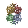
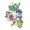
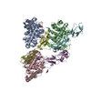
| Title | D-Hydantoinase (Dihydropyrimidinase) from Thermus sp. in space group C2221 | ||||||
 Components Components | HYDANTOINASE | ||||||
 Keywords Keywords | HYDROLASE / HYDANTOINASE / DIHYDROPYRIMIDINASE / CYCLIC AMIDASE | ||||||
| Function / homology |  Function and homology information Function and homology informationhydrolase activity, acting on carbon-nitrogen (but not peptide) bonds, in cyclic amides / metal ion binding / cytosol Similarity search - Function | ||||||
| Biological species |   THERMUS SP. (bacteria) THERMUS SP. (bacteria) | ||||||
| Method |  X-RAY DIFFRACTION / SYNCHROTRON / MAD / Resolution: 1.295 Å X-RAY DIFFRACTION / SYNCHROTRON / MAD / Resolution: 1.295 Å | ||||||
 Authors Authors | Abendroth, J. / Niefind, K. / Schomburg, D. | ||||||
 Citation Citation | Journal: J.Mol.Biol. / Year: 2002 Title: X-Ray Structure of a Dihydropyrimidinase from Thermus Sp. At 1.3 A Resolution Authors: Abendroth, J. / Niefind, K. / Schomburg, D. #1: Journal: Acta Crystallogr.,Sect.D / Year: 2000 Title: Crystallization, Preliminary X-Ray Analysis of a Native and Selenomethionine D-Hydantoinase from Thermus Sp Authors: Abendroth, J. / Niefind, K. / Schomburg, D. | ||||||
| History |
| ||||||
| Remark 700 | SHEET THE SHEET STRUCTURE OF THIS MOLECULE IS BIFURCATED. IN ORDER TO REPRESENT THIS FEATURE IN ... SHEET THE SHEET STRUCTURE OF THIS MOLECULE IS BIFURCATED. IN ORDER TO REPRESENT THIS FEATURE IN THE SHEET RECORDS BELOW, TWO SHEETS ARE DEFINED. |
- Structure visualization
| Structure viewer | Molecule: MolmilJmol/JSmol |
|---|
- Downloads & links
-Download
| PDBx/mmCIF format | 1gkp.cif.gz | 1.2 MB | Display | PDBx/mmCIF format |
|---|---|---|---|---|
| PDB format | pdb1gkp.ent.gz | 998.7 KB | Display | PDB format |
| PDBx/mmJSON format | 1gkp.json.gz | Tree view | PDBx/mmJSON format | |
| Others |  Other downloads Other downloads |
-Validation report
| Arichive directory | https://data.pdbj.org/pub/pdb/validation_reports/gk/1gkpftp://data.pdbj.org/pub/pdb/validation_reports/gk/1gkp | HTTPS FTP |
|---|
-Related structure data


-Links
 PDBj
PDBj

- Assembly
| Deposited unit | 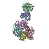
| ||||||||||||||||||||||||
|---|---|---|---|---|---|---|---|---|---|---|---|---|---|---|---|---|---|---|---|---|---|---|---|---|---|
| 1 | 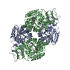
| ||||||||||||||||||||||||
| 2 | 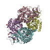
| ||||||||||||||||||||||||
| Unit cell |
| ||||||||||||||||||||||||
| Noncrystallographic symmetry (NCS) | NCS oper:
|
-Components
| #1: Protein | Mass: 50781.422 Da / Num. of mol.: 6 Source method: isolated from a genetically manipulated source Details: KCX IS A NZ-CARBOXYLATED LYSINE / Source: (gene. exp.) THERMUS SP. (bacteria) / Production host: #2: Chemical | ChemComp-ZN /   Mass: 65.409 Da / Num. of mol.: 12 / Source method: obtained synthetically / Formula: Zn Mass: 65.409 Da / Num. of mol.: 12 / Source method: obtained synthetically / Formula: Zn#3: Chemical | ChemComp-SO4 /   Mass: 96.063 Da / Num. of mol.: 9 / Source method: obtained synthetically / Formula: SO4 Mass: 96.063 Da / Num. of mol.: 9 / Source method: obtained synthetically / Formula: SO4#4: Chemical |   Mass: 238.305 Da / Num. of mol.: 2 / Source method: obtained synthetically / Formula: C8H18N2O4S / Comment: pH buffer*YM Mass: 238.305 Da / Num. of mol.: 2 / Source method: obtained synthetically / Formula: C8H18N2O4S / Comment: pH buffer*YM#5: Water | ChemComp-HOH / |  Mass: 18.015 Da / Num. of mol.: 3716 / Source method: isolated from a natural source / Formula: H2O Mass: 18.015 Da / Num. of mol.: 3716 / Source method: isolated from a natural source / Formula: H2OHas protein modification | Y | |
|---|
-Experimental details
-Experiment
| Experiment | Method: X-RAY DIFFRACTION / Number of used crystals: 1 |
|---|
- Sample preparation
| Crystal | Density Matthews: 2.3 Å3/Da / Density % sol: 41 % Description: MAD DATA TO 3AA, INITIAL MODEL FROM ARP AT 1.7AA | ||||||||||||||||||||||||||||||||||||
|---|---|---|---|---|---|---|---|---|---|---|---|---|---|---|---|---|---|---|---|---|---|---|---|---|---|---|---|---|---|---|---|---|---|---|---|---|---|
| Crystal grow | pH: 7.5 Details: 1.65 MM AMMONIUM SULPHATE, 100 MM HEPES/NAOH PH 7.5, 5% (V/V) PEG 400, CRYO-BUFFER: 2 M LITHIUM SULPHATE, 100 MM HEPES/NAOH PH 7.5, 5% (V/V) PEG 400 | ||||||||||||||||||||||||||||||||||||
| Crystal grow | *PLUS Method: vapor diffusion, sitting drop | ||||||||||||||||||||||||||||||||||||
| Components of the solutions | *PLUS
|
-Data collection
| Diffraction | Mean temperature: 100 K |
|---|---|
| Diffraction source | Source: SYNCHROTRON / Site: EMBL/DESY, HAMBURG  / Beamline: BW7B / Wavelength: 0.842 / Beamline: BW7B / Wavelength: 0.842 |
| Detector | Type: MAR scanner 345 mm plate / Detector: IMAGE PLATE / Date: Aug 24, 2000 |
| Radiation | Monochromator: TRIANGULAR MONOCHROMATOR / Protocol: SINGLE WAVELENGTH / Monochromatic (M) / Laue (L): M / Scattering type: x-ray |
| Radiation wavelength | Wavelength: 0.842 Å / Relative weight: 1 |
| Reflection | Resolution: 1.3→50 Å / Num. obs: 693388 / % possible obs: 99.7 % / Redundancy: 4.3 % / Rmerge(I) obs: 0.031 / Net I/σ(I): 17.7 |
| Reflection shell | Resolution: 1.3→1.33 Å / Redundancy: 3.7 % / Rmerge(I) obs: 0.425 / Mean I/σ(I) obs: 2.4 / % possible all: 98 |
| Reflection | *PLUS Lowest resolution: 50 Å / Num. measured all: 2976914 |
| Reflection shell | *PLUS % possible obs: 98 % |
- Processing
| Software |
| ||||||||||||||||||||||||
|---|---|---|---|---|---|---|---|---|---|---|---|---|---|---|---|---|---|---|---|---|---|---|---|---|---|
| Refinement | Method to determine structure: MAD / Resolution: 1.295→50 Å / SU B: 1.996 / SU ML: 0.045 / Cross valid method: THROUGHOUT / ESU R: 0.047 / ESU R Free: 0.046 / Details: NONE
| ||||||||||||||||||||||||
| Refinement step | Cycle: LAST / Resolution: 1.295→50 Å
| ||||||||||||||||||||||||
| Refinement | *PLUS Highest resolution: 1.3 Å / Lowest resolution: 50 Å / Rfactor Rfree: 0.184 / Rfactor Rwork: 0.153 | ||||||||||||||||||||||||
| Solvent computation | *PLUS | ||||||||||||||||||||||||
| Displacement parameters | *PLUS | ||||||||||||||||||||||||
| Refine LS restraints | *PLUS
|
