 Movie
Movie Controller

[English] 日本語
 Yorodumi
- PDB-1gkq: D-Hydantoinase (Dihydropyrimidinase) from Thermus sp. in space gr... -
+ Open data

- Basic information
| Entry | Database: PDB / ID: 1gkq | ||||||
|---|---|---|---|---|---|---|---|
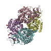
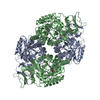
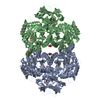
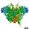
| Title | D-Hydantoinase (Dihydropyrimidinase) from Thermus sp. in space group P212121 | ||||||
 Components Components | HYDANTOINASE | ||||||
 Keywords Keywords | HYDROLASE / HYDANTOINASE / DIHYDROPYRIMIDINASE / CYCLIC AMIDASE | ||||||
| Function / homology |  Function and homology information Function and homology informationhydrolase activity, acting on carbon-nitrogen (but not peptide) bonds, in cyclic amides / metal ion binding / cytosol Similarity search - Function | ||||||
| Biological species |   THERMUS SP. (bacteria) THERMUS SP. (bacteria) | ||||||
| Method |  X-RAY DIFFRACTION / MOLECULAR REPLACEMENT / Resolution: 2.6 Å X-RAY DIFFRACTION / MOLECULAR REPLACEMENT / Resolution: 2.6 Å | ||||||
 Authors Authors | Abendroth, J. / Niefind, K. / Schomburg, D. | ||||||
 Citation Citation | Journal: J.Mol.Biol. / Year: 2002 Title: X-Ray Structure of a Dihydropyrimidinase from Thermus Sp. At 1.3 A Resolution Authors: Abendroth, J. / Niefind, K. / Schomburg, D. | ||||||
| History |
| ||||||
| Remark 700 | SHEET THE SHEET STRUCTURE OF THIS MOLECULE IS BIFURCATED. IN ORDER TO REPRESENT THIS FEATURE IN ... SHEET THE SHEET STRUCTURE OF THIS MOLECULE IS BIFURCATED. IN ORDER TO REPRESENT THIS FEATURE IN THE SHEET RECORDS BELOW, TWO SHEETS ARE DEFINED. |
- Structure visualization
| Structure viewer | Molecule: MolmilJmol/JSmol |
|---|
- Downloads & links
-Download
| PDBx/mmCIF format | 1gkq.cif.gz | 365.2 KB | Display | PDBx/mmCIF format |
|---|---|---|---|---|
| PDB format | pdb1gkq.ent.gz | 299 KB | Display | PDB format |
| PDBx/mmJSON format | 1gkq.json.gz | Tree view | PDBx/mmJSON format | |
| Others |  Other downloads Other downloads |
-Validation report
| Arichive directory | https://data.pdbj.org/pub/pdb/validation_reports/gk/1gkqftp://data.pdbj.org/pub/pdb/validation_reports/gk/1gkq | HTTPS FTP |
|---|
-Related structure data
| Related structure data |  1gkpSC S: Starting model for refinement C: citing same article ( |
|---|---|
| Similar structure data |


-Links
 PDBj
PDBj

- Assembly
| Deposited unit | 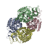
| ||||||||||||||||
|---|---|---|---|---|---|---|---|---|---|---|---|---|---|---|---|---|---|
| 1 |
| ||||||||||||||||
| Unit cell |
| ||||||||||||||||
| Noncrystallographic symmetry (NCS) | NCS oper:
|
-Components
| #1: Protein | Mass: 50781.422 Da / Num. of mol.: 4 Source method: isolated from a genetically manipulated source Details: KCX IS A NZ-CARBOXYLATED LYSINE / Source: (gene. exp.) THERMUS SP. (bacteria) / Description: RECOMBINANTLY EXPRESSED / Production host: #2: Chemical | ChemComp-ZN /   Mass: 65.409 Da / Num. of mol.: 8 / Source method: obtained synthetically / Formula: Zn Mass: 65.409 Da / Num. of mol.: 8 / Source method: obtained synthetically / Formula: Zn#3: Water | ChemComp-HOH / |  Mass: 18.015 Da / Num. of mol.: 489 / Source method: isolated from a natural source / Formula: H2O Mass: 18.015 Da / Num. of mol.: 489 / Source method: isolated from a natural source / Formula: H2O |
|---|
-Experimental details
-Experiment
| Experiment | Method: X-RAY DIFFRACTION / Number of used crystals: 1 |
|---|
- Sample preparation
| Crystal | Density Matthews: 2.8 Å3/Da / Density % sol: 61 % | ||||||||||||||||||||||||||||||
|---|---|---|---|---|---|---|---|---|---|---|---|---|---|---|---|---|---|---|---|---|---|---|---|---|---|---|---|---|---|---|---|
| Crystal grow | pH: 6.5 Details: 30% (W/V) PEG 6000, 100 MM CACODYLATE/HCL PH 6.5, 200 MM SODIUM ACETATE | ||||||||||||||||||||||||||||||
| Crystal grow | *PLUS Method: vapor diffusion, sitting drop | ||||||||||||||||||||||||||||||
| Components of the solutions | *PLUS
|
-Data collection
| Diffraction | Mean temperature: 283 K |
|---|---|
| Diffraction source | Source: ROTATING ANODE / Type: ENRAF-NONIUS FR591 / Wavelength: 1.5418 |
| Detector | Type: MAC SCIENCE DIP-2030H / Detector: IMAGE PLATE / Date: Nov 6, 1997 |
| Radiation | Monochromator: NI FITER / Protocol: SINGLE WAVELENGTH / Monochromatic (M) / Laue (L): M / Scattering type: x-ray |
| Radiation wavelength | Wavelength: 1.5418 Å / Relative weight: 1 |
| Reflection | Resolution: 2.6→25 Å / Num. obs: 70394 / % possible obs: 100 % / Redundancy: 5.9 % / Biso Wilson estimate: 38.8 Å2 / Rmerge(I) obs: 0.117 / Net I/σ(I): 12 |
| Reflection shell | Resolution: 2.6→2.69 Å / Redundancy: 5.7 % / Rmerge(I) obs: 0.363 / Mean I/σ(I) obs: 4.9 / % possible all: 99.9 |
| Reflection | *PLUS Highest resolution: 2.6 Å / Lowest resolution: 25 Å / % possible obs: 99.9 % / Num. measured all: 416020 |
| Reflection shell | *PLUS % possible obs: 99.9 % |
- Processing
| Software |
| ||||||||||||||||||||||||||||||||||||||||||||||||||||||||||||
|---|---|---|---|---|---|---|---|---|---|---|---|---|---|---|---|---|---|---|---|---|---|---|---|---|---|---|---|---|---|---|---|---|---|---|---|---|---|---|---|---|---|---|---|---|---|---|---|---|---|---|---|---|---|---|---|---|---|---|---|---|---|
| Refinement | Method to determine structure: MOLECULAR REPLACEMENT Starting model: 1GKP Resolution: 2.6→25 Å / Rfactor Rfree error: 0.0035 / Data cutoff high absF: 10000 / Cross valid method: THROUGHOUT / σ(F): 0
| ||||||||||||||||||||||||||||||||||||||||||||||||||||||||||||
| Solvent computation | Bsol: 42.4 Å2 / ksol: 0.355 e/Å3 | ||||||||||||||||||||||||||||||||||||||||||||||||||||||||||||
| Displacement parameters | Biso mean: 28.3 Å2
| ||||||||||||||||||||||||||||||||||||||||||||||||||||||||||||
| Refine analyze |
| ||||||||||||||||||||||||||||||||||||||||||||||||||||||||||||
| Refinement step | Cycle: LAST / Resolution: 2.6→25 Å
| ||||||||||||||||||||||||||||||||||||||||||||||||||||||||||||
| Refine LS restraints |
| ||||||||||||||||||||||||||||||||||||||||||||||||||||||||||||
| Refine LS restraints NCS | NCS model details: RESTRAINTS / Rms dev position: 300 Å | ||||||||||||||||||||||||||||||||||||||||||||||||||||||||||||
| LS refinement shell | Resolution: 2.6→2.69 Å / Rfactor Rfree error: 0.016 / Total num. of bins used: 10
| ||||||||||||||||||||||||||||||||||||||||||||||||||||||||||||
| Refinement | *PLUS Rfactor Rfree: 0.211 / Rfactor Rwork: 0.188 | ||||||||||||||||||||||||||||||||||||||||||||||||||||||||||||
| Solvent computation | *PLUS | ||||||||||||||||||||||||||||||||||||||||||||||||||||||||||||
| Displacement parameters | *PLUS | ||||||||||||||||||||||||||||||||||||||||||||||||||||||||||||
| Refine LS restraints | *PLUS
|
