+ データを開く
- 基本情報
基本情報
| 登録情報 | データベース: PDB / ID: 6u7k | |||||||||
|---|---|---|---|---|---|---|---|---|---|---|
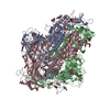
| タイトル | Prefusion structure of PEDV spike | |||||||||
 要素 要素 | Spike glycoprotein | |||||||||
 キーワード キーワード | VIRAL PROTEIN / PEDV / Spike / Coronavirus / Fusion Protein | |||||||||
| 機能・相同性 |  機能・相同性情報 機能・相同性情報host cell endoplasmic reticulum-Golgi intermediate compartment membrane / receptor-mediated virion attachment to host cell / endocytosis involved in viral entry into host cell / fusion of virus membrane with host plasma membrane / fusion of virus membrane with host endosome membrane / viral envelope / virion membrane / membrane 類似検索 - 分子機能 | |||||||||
| 生物種 |  Porcine epidemic diarrhea virus (ブタ流行性下痢ウイルス) Porcine epidemic diarrhea virus (ブタ流行性下痢ウイルス) | |||||||||
| 手法 | 電子顕微鏡法 / 単粒子再構成法 / クライオ電子顕微鏡法 / 解像度: 3.14 Å | |||||||||
 データ登録者 データ登録者 | Wrapp, D. / McLellan, J.S. | |||||||||
| 資金援助 |  米国, 1件 米国, 1件
| |||||||||
 引用 引用 |  ジャーナル: J Virol / 年: 2019 ジャーナル: J Virol / 年: 2019タイトル: The 3.1-Angstrom Cryo-electron Microscopy Structure of the Porcine Epidemic Diarrhea Virus Spike Protein in the Prefusion Conformation. 著者: Daniel Wrapp / Jason S McLellan /  要旨: Porcine epidemic diarrhea virus (PEDV) is an alphacoronavirus that has a significant agricultural and economic impact due to the high mortality rate associated with infection of neonatal piglets. ...Porcine epidemic diarrhea virus (PEDV) is an alphacoronavirus that has a significant agricultural and economic impact due to the high mortality rate associated with infection of neonatal piglets. Like other coronaviruses, PEDV makes use of a large, trimeric spike (S) glycoprotein to mediate membrane fusion and gain entry into host cells. Despite the importance of the spike protein in viral entry and host immune responses, high-resolution structural information concerning this large macromolecular machine has been difficult to obtain. Here, we report the cryo-electron microscopy structure of the PEDV S protein in the prefusion conformation at a resolution of 3.1 Å. Our studies revealed that the sialic acid-binding domain at the N terminus of the S1 subunit has an orientation that is substantially different from that observed in the previously determined spike structure from human alphacoronavirus NL63. We also observed dissociated S1 subunit trimers wherein the putative receptor-binding domains exist in a conformation differing from that observed in the intact spike proteins, suggesting that the PEDV receptor-binding domain undergoes conformational rearrangements akin to those that have been described in the related betacoronaviruses. Collectively, these data provide new insights into the biological processes that mediate alphacoronavirus attachment, receptor engagement, and fusion triggering while also identifying a source of conformational heterogeneity that could be manipulated to improve PEDV vaccine antigens. Coronavirus spike proteins are large, densely glycosylated macromolecular machines that mediate receptor binding and membrane fusion to facilitate entry into host cells. This report describes the atomic-resolution structure of the spike protein from porcine epidemic diarrhea virus, a pathogenic alphacoronavirus that causes severe agricultural damage. The structure reveals a novel position for the sialic acid-binding attachment domain in the intact spike. We also observed shed fusion-suppressive capping subunits that displayed the putative receptor-binding domain in an accessible conformation. These observations provide a basis for understanding the molecular mechanisms that drive the earliest stages of alphacoronavirus infection and will inform future efforts to rationally design vaccines. | |||||||||
| 履歴 |
|
- 構造の表示
構造の表示
| ムービー |
 ムービービューア ムービービューア |
|---|---|
| 構造ビューア | 分子:  Molmil Molmil Jmol/JSmol Jmol/JSmol |
- ダウンロードとリンク
- ダウンロード
| PDBx/mmCIF形式 |  6u7k.cif.gz 6u7k.cif.gz | 583.9 KB | 表示 |  PDBx/mmCIF形式 PDBx/mmCIF形式 |
|---|---|---|---|---|
| PDB形式 |  pdb6u7k.ent.gz pdb6u7k.ent.gz | 472.1 KB | 表示 |  PDB形式 PDB形式 |
| PDBx/mmJSON形式 |  6u7k.json.gz 6u7k.json.gz | ツリー表示 |  PDBx/mmJSON形式 PDBx/mmJSON形式 | |
| その他 |  その他のダウンロード その他のダウンロード |
-検証レポート
| 文書・要旨 |  6u7k_validation.pdf.gz 6u7k_validation.pdf.gz | 2.4 MB | 表示 |  wwPDB検証レポート wwPDB検証レポート |
|---|---|---|---|---|
| 文書・詳細版 |  6u7k_full_validation.pdf.gz 6u7k_full_validation.pdf.gz | 2.5 MB | 表示 | |
| XML形式データ |  6u7k_validation.xml.gz 6u7k_validation.xml.gz | 115 KB | 表示 | |
| CIF形式データ |  6u7k_validation.cif.gz 6u7k_validation.cif.gz | 167.6 KB | 表示 | |
| アーカイブディレクトリ |  https://data.pdbj.org/pub/pdb/validation_reports/u7/6u7k https://data.pdbj.org/pub/pdb/validation_reports/u7/6u7k ftp://data.pdbj.org/pub/pdb/validation_reports/u7/6u7k ftp://data.pdbj.org/pub/pdb/validation_reports/u7/6u7k | HTTPS FTP |
-関連構造データ
- リンク
リンク
- 集合体
集合体
| 登録構造単位 | 
|
|---|---|
| 1 |
|
- 要素
要素
| #1: タンパク質 | 分子量: 152906.906 Da / 分子数: 3 / 由来タイプ: 組換発現 由来: (組換発現)  Porcine epidemic diarrhea virus (strain CV777) (ウイルス) Porcine epidemic diarrhea virus (strain CV777) (ウイルス)株: CV777 / 遺伝子: S, 2 / 発現宿主:  Homo sapiens (ヒト) / 参照: UniProt: Q91AV1 Homo sapiens (ヒト) / 参照: UniProt: Q91AV1#2: 多糖 | #3: 多糖 | 2-acetamido-2-deoxy-beta-D-glucopyranose-(1-4)-2-acetamido-2-deoxy-beta-D-glucopyranose #4: 糖 | ChemComp-NAG / 研究の焦点であるリガンドがあるか | N | Has protein modification | Y | |
|---|
-実験情報
-実験
| 実験 | 手法: 電子顕微鏡法 |
|---|---|
| EM実験 | 試料の集合状態: PARTICLE / 3次元再構成法: 単粒子再構成法 |
- 試料調製
試料調製
| 構成要素 | 名称: Homotrimeric complex of PEDV spike / タイプ: COMPLEX / Entity ID: #1 / 由来: RECOMBINANT | |||||||||||||||||||||||||
|---|---|---|---|---|---|---|---|---|---|---|---|---|---|---|---|---|---|---|---|---|---|---|---|---|---|---|
| 分子量 | 値: 0.452 MDa / 実験値: NO | |||||||||||||||||||||||||
| 由来(天然) | 生物種:  Porcine epidemic diarrhea virus CV777 (ウイルス) Porcine epidemic diarrhea virus CV777 (ウイルス) | |||||||||||||||||||||||||
| 由来(組換発現) | 生物種:  Homo sapiens (ヒト) Homo sapiens (ヒト) | |||||||||||||||||||||||||
| ウイルスについての詳細 | 単離: STRAIN | |||||||||||||||||||||||||
| 緩衝液 | pH: 8 | |||||||||||||||||||||||||
| 緩衝液成分 |
| |||||||||||||||||||||||||
| 試料 | 濃度: 0.4 mg/ml / 包埋: NO / シャドウイング: NO / 染色: NO / 凍結: YES | |||||||||||||||||||||||||
| 試料支持 | グリッドの材料: COPPER / グリッドのサイズ: 400 divisions/in. / グリッドのタイプ: C-flat-2/2 | |||||||||||||||||||||||||
| 急速凍結 | 装置: FEI VITROBOT MARK IV / 凍結剤: ETHANE / 湿度: 100 % / 凍結前の試料温度: 277.15 K / 詳細: Blot for (6) seconds before plunging |
- 電子顕微鏡撮影
| 実験機器 |  モデル: Titan Krios / 画像提供: FEI Company |
|---|---|
| 顕微鏡 | モデル: FEI TITAN KRIOS |
| 電子銃 | 電子線源:  FIELD EMISSION GUN / 加速電圧: 300 kV / 照射モード: FLOOD BEAM FIELD EMISSION GUN / 加速電圧: 300 kV / 照射モード: FLOOD BEAM |
| 電子レンズ | モード: BRIGHT FIELD / Cs: 2.7 mm / C2レンズ絞り径: 100 µm |
| 試料ホルダ | 凍結剤: NITROGEN 試料ホルダーモデル: FEI TITAN KRIOS AUTOGRID HOLDER |
| 撮影 | 電子線照射量: 48 e/Å2 / 検出モード: COUNTING フィルム・検出器のモデル: GATAN K2 SUMMIT (4k x 4k) |
- 解析
解析
| EMソフトウェア |
| ||||||||||||||||||||||||||||
|---|---|---|---|---|---|---|---|---|---|---|---|---|---|---|---|---|---|---|---|---|---|---|---|---|---|---|---|---|---|
| CTF補正 | タイプ: PHASE FLIPPING AND AMPLITUDE CORRECTION | ||||||||||||||||||||||||||||
| 対称性 | 点対称性: C3 (3回回転対称) | ||||||||||||||||||||||||||||
| 3次元再構成 | 解像度: 3.14 Å / 解像度の算出法: FSC 0.143 CUT-OFF / 粒子像の数: 112655 / アルゴリズム: FOURIER SPACE 詳細: Final reconstruction calculated using non-uniform 3D refinement クラス平均像の数: 1 / 対称性のタイプ: POINT |
 ムービー
ムービー コントローラー


 PDBj
PDBj

