National Institutes of Health/National Institute Of Allergy and Infectious Diseases (NIH/NIAID)
R01-AI127521
United States
Citation
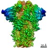
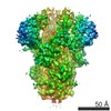
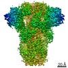
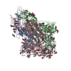
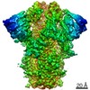
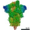
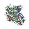
Journal: J Virol / Year: 2019 Title: The 3.1-Angstrom Cryo-electron Microscopy Structure of the Porcine Epidemic Diarrhea Virus Spike Protein in the Prefusion Conformation. Authors: Daniel Wrapp / Jason S McLellan / Abstract: Porcine epidemic diarrhea virus (PEDV) is an alphacoronavirus that has a significant agricultural and economic impact due to the high mortality rate associated with infection of neonatal piglets. ...Porcine epidemic diarrhea virus (PEDV) is an alphacoronavirus that has a significant agricultural and economic impact due to the high mortality rate associated with infection of neonatal piglets. Like other coronaviruses, PEDV makes use of a large, trimeric spike (S) glycoprotein to mediate membrane fusion and gain entry into host cells. Despite the importance of the spike protein in viral entry and host immune responses, high-resolution structural information concerning this large macromolecular machine has been difficult to obtain. Here, we report the cryo-electron microscopy structure of the PEDV S protein in the prefusion conformation at a resolution of 3.1 Å. Our studies revealed that the sialic acid-binding domain at the N terminus of the S1 subunit has an orientation that is substantially different from that observed in the previously determined spike structure from human alphacoronavirus NL63. We also observed dissociated S1 subunit trimers wherein the putative receptor-binding domains exist in a conformation differing from that observed in the intact spike proteins, suggesting that the PEDV receptor-binding domain undergoes conformational rearrangements akin to those that have been described in the related betacoronaviruses. Collectively, these data provide new insights into the biological processes that mediate alphacoronavirus attachment, receptor engagement, and fusion triggering while also identifying a source of conformational heterogeneity that could be manipulated to improve PEDV vaccine antigens. Coronavirus spike proteins are large, densely glycosylated macromolecular machines that mediate receptor binding and membrane fusion to facilitate entry into host cells. This report describes the atomic-resolution structure of the spike protein from porcine epidemic diarrhea virus, a pathogenic alphacoronavirus that causes severe agricultural damage. The structure reveals a novel position for the sialic acid-binding attachment domain in the intact spike. We also observed shed fusion-suppressive capping subunits that displayed the putative receptor-binding domain in an accessible conformation. These observations provide a basis for understanding the molecular mechanisms that drive the earliest stages of alphacoronavirus infection and will inform future efforts to rationally design vaccines.
History
Deposition
Sep 3, 2019
-
Header (metadata) release
Sep 18, 2019
-
Map release
Sep 18, 2019
-
Update
Oct 23, 2024
-
Current status
Oct 23, 2024
Processing site: RCSB / Status: Released
-
Structure visualization
Movie
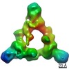
Surface view with section colored by density value
Model: C-flat-2/2 / Material: COPPER / Mesh: 400 / Support film - Material: CARBON / Support film - topology: HOLEY ARRAY / Pretreatment - Type: PLASMA CLEANING / Pretreatment - Time: 30 sec. / Pretreatment - Atmosphere: OTHER
Vitrification
Cryogen name: ETHANE / Chamber humidity: 100 % / Chamber temperature: 277.15 K / Instrument: FEI VITROBOT MARK IV / Details: Blot for (6) seconds before plunging.
-
Electron microscopy
Microscope
FEI TITAN KRIOS
Image recording
Film or detector model: GATAN K2 SUMMIT (4k x 4k) / Detector mode: COUNTING / Average electron dose: 48.0 e/Å2
Electron beam
Acceleration voltage: 300 kV / Electron source: FIELD EMISSION GUN
Electron optics
C2 aperture diameter: 100.0 µm / Illumination mode: FLOOD BEAM / Imaging mode: BRIGHT FIELD / Cs: 2.7 mm
In the structure databanks used in Yorodumi, some data are registered as the other names, "COVID-19 virus" and "2019-nCoV". Here are the details of the virus and the list of structure data.
Jan 31, 2019. EMDB accession codes are about to change! (news from PDBe EMDB page)
EMDB accession codes are about to change! (news from PDBe EMDB page)
The allocation of 4 digits for EMDB accession codes will soon come to an end. Whilst these codes will remain in use, new EMDB accession codes will include an additional digit and will expand incrementally as the available range of codes is exhausted. The current 4-digit format prefixed with “EMD-” (i.e. EMD-XXXX) will advance to a 5-digit format (i.e. EMD-XXXXX), and so on. It is currently estimated that the 4-digit codes will be depleted around Spring 2019, at which point the 5-digit format will come into force.
The EM Navigator/Yorodumi systems omit the EMD- prefix.
Related info.:Q: What is EMD? / ID/Accession-code notation in Yorodumi/EM Navigator
Yorodumi is a browser for structure data from EMDB, PDB, SASBDB, etc.
This page is also the successor to EM Navigator detail page, and also detail information page/front-end page for Omokage search.
The word "yorodu" (or yorozu) is an old Japanese word meaning "ten thousand". "mi" (miru) is to see.
Related info.:EMDB / PDB / SASBDB / Comparison of 3 databanks / Yorodumi Search / Aug 31, 2016. New EM Navigator & Yorodumi / Yorodumi Papers / Jmol/JSmol / Function and homology information / Changes in new EM Navigator and Yorodumi
 Movie
Movie Controller


 Open data
 Basic information
 Map data
 Sample
 Keywords
 Function and homology information
 Porcine epidemic diarrhea virus CV777 /
  Authors
 United States, 1 items
  Citation
 Structure visualization
 Downloads & links
 emd_20672.png
 http://ftp.pdbj.org/pub/emdb/structures/EMD-20672
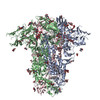

 Z (Sec.)
 Y (Row.)
 X (Col.)


 Sample components
 Homo sapiens (human)
 Processing
 Electron microscopy
 FIELD EMISSION GUN

