 Movie
Movie Controller


+ Open data

- Basic information
| Entry | Database: PDB / ID: 6u7k | |||||||||
|---|---|---|---|---|---|---|---|---|---|---|
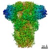
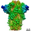
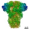
| Title | Prefusion structure of PEDV spike | |||||||||
 Components Components | Spike glycoprotein | |||||||||
 Keywords Keywords | VIRAL PROTEIN / PEDV / Spike / Coronavirus / Fusion Protein | |||||||||
| Function / homology |  Function and homology information Function and homology informationhost cell endoplasmic reticulum-Golgi intermediate compartment membrane / receptor-mediated virion attachment to host cell / endocytosis involved in viral entry into host cell / fusion of virus membrane with host plasma membrane / fusion of virus membrane with host endosome membrane / viral envelope / virion membrane / membrane Similarity search - Function | |||||||||
| Biological species |  Porcine epidemic diarrhea virus Porcine epidemic diarrhea virus | |||||||||
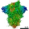
| Method | ELECTRON MICROSCOPY / single particle reconstruction / cryo EM / Resolution: 3.14 Å | |||||||||
 Authors Authors | Wrapp, D. / McLellan, J.S. | |||||||||
| Funding support |  United States, 1items United States, 1items
| |||||||||
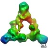
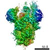
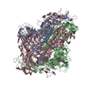
 Citation Citation | Journal: J Virol / Year: 2019 Title: The 3.1-Angstrom Cryo-electron Microscopy Structure of the Porcine Epidemic Diarrhea Virus Spike Protein in the Prefusion Conformation. Authors: Daniel Wrapp / Jason S McLellan / Abstract: Porcine epidemic diarrhea virus (PEDV) is an alphacoronavirus that has a significant agricultural and economic impact due to the high mortality rate associated with infection of neonatal piglets. ...Porcine epidemic diarrhea virus (PEDV) is an alphacoronavirus that has a significant agricultural and economic impact due to the high mortality rate associated with infection of neonatal piglets. Like other coronaviruses, PEDV makes use of a large, trimeric spike (S) glycoprotein to mediate membrane fusion and gain entry into host cells. Despite the importance of the spike protein in viral entry and host immune responses, high-resolution structural information concerning this large macromolecular machine has been difficult to obtain. Here, we report the cryo-electron microscopy structure of the PEDV S protein in the prefusion conformation at a resolution of 3.1 Å. Our studies revealed that the sialic acid-binding domain at the N terminus of the S1 subunit has an orientation that is substantially different from that observed in the previously determined spike structure from human alphacoronavirus NL63. We also observed dissociated S1 subunit trimers wherein the putative receptor-binding domains exist in a conformation differing from that observed in the intact spike proteins, suggesting that the PEDV receptor-binding domain undergoes conformational rearrangements akin to those that have been described in the related betacoronaviruses. Collectively, these data provide new insights into the biological processes that mediate alphacoronavirus attachment, receptor engagement, and fusion triggering while also identifying a source of conformational heterogeneity that could be manipulated to improve PEDV vaccine antigens. Coronavirus spike proteins are large, densely glycosylated macromolecular machines that mediate receptor binding and membrane fusion to facilitate entry into host cells. This report describes the atomic-resolution structure of the spike protein from porcine epidemic diarrhea virus, a pathogenic alphacoronavirus that causes severe agricultural damage. The structure reveals a novel position for the sialic acid-binding attachment domain in the intact spike. We also observed shed fusion-suppressive capping subunits that displayed the putative receptor-binding domain in an accessible conformation. These observations provide a basis for understanding the molecular mechanisms that drive the earliest stages of alphacoronavirus infection and will inform future efforts to rationally design vaccines. | |||||||||
| History |
|
- Structure visualization
| Movie |
Movie viewer |
|---|---|
| Structure viewer | Molecule: MolmilJmol/JSmol |
- Downloads & links
-Download
| PDBx/mmCIF format | 6u7k.cif.gz | 583.9 KB | Display | PDBx/mmCIF format |
|---|---|---|---|---|
| PDB format | pdb6u7k.ent.gz | 472.1 KB | Display | PDB format |
| PDBx/mmJSON format | 6u7k.json.gz | Tree view | PDBx/mmJSON format | |
| Others |  Other downloads Other downloads |
-Validation report
| Arichive directory | https://data.pdbj.org/pub/pdb/validation_reports/u7/6u7kftp://data.pdbj.org/pub/pdb/validation_reports/u7/6u7k | HTTPS FTP |
|---|
-Related structure data
| Related structure data | 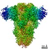 20672MC M: map data used to model this data C: citing same article ( |
|---|---|
| Similar structure data |


-Links
 PDBj
PDBj- Assembly
| Deposited unit | 
|
|---|---|
| 1 |
|
-Components
| #1: Protein | Mass: 152906.906 Da / Num. of mol.: 3 Source method: isolated from a genetically manipulated source Source: (gene. exp.) Porcine epidemic diarrhea virus (strain CV777)Strain: CV777 / Gene: S, 2 / Production host:  Homo sapiens (human) / References: UniProt: Q91AV1 Homo sapiens (human) / References: UniProt: Q91AV1#2: Polysaccharide | Source method: isolated from a genetically manipulated source #3: Polysaccharide | 2-acetamido-2-deoxy-beta-D-glucopyranose-(1-4)-2-acetamido-2-deoxy-beta-D-glucopyranose Source method: isolated from a genetically manipulated source #4: Sugar | ChemComp-NAG /   Type: D-saccharide, beta linking / Mass: 221.208 Da / Num. of mol.: 39 Type: D-saccharide, beta linking / Mass: 221.208 Da / Num. of mol.: 39Source method: isolated from a genetically manipulated source Formula: C8H15NO6 Has ligand of interest | N | Has protein modification | Y | |
|---|
-Experimental details
-Experiment
| Experiment | Method: ELECTRON MICROSCOPY |
|---|---|
| EM experiment | Aggregation state: PARTICLE / 3D reconstruction method: single particle reconstruction |
- Sample preparation
| Component | Name: Homotrimeric complex of PEDV spike / Type: COMPLEX / Entity ID: #1 / Source: RECOMBINANT | |||||||||||||||||||||||||
|---|---|---|---|---|---|---|---|---|---|---|---|---|---|---|---|---|---|---|---|---|---|---|---|---|---|---|
| Molecular weight | Value: 0.452 MDa / Experimental value: NO | |||||||||||||||||||||||||
| Source (natural) | Organism: Porcine epidemic diarrhea virus CV777 | |||||||||||||||||||||||||
| Source (recombinant) | Organism: Homo sapiens (human) | |||||||||||||||||||||||||
| Details of virus | Isolate: STRAIN | |||||||||||||||||||||||||
| Buffer solution | pH: 8 | |||||||||||||||||||||||||
| Buffer component |
| |||||||||||||||||||||||||
| Specimen | Conc.: 0.4 mg/ml / Embedding applied: NO / Shadowing applied: NO / Staining applied: NO / Vitrification applied: YES | |||||||||||||||||||||||||
| Specimen support | Grid material: COPPER / Grid mesh size: 400 divisions/in. / Grid type: C-flat-2/2 | |||||||||||||||||||||||||
| Vitrification | Instrument: FEI VITROBOT MARK IV / Cryogen name: ETHANE / Humidity: 100 % / Chamber temperature: 277.15 K / Details: Blot for (6) seconds before plunging |
- Electron microscopy imaging
| Experimental equipment |  Model: Titan Krios / Image courtesy: FEI Company |
|---|---|
| Microscopy | Model: FEI TITAN KRIOS |
| Electron gun | Electron source:  FIELD EMISSION GUN / Accelerating voltage: 300 kV / Illumination mode: FLOOD BEAM FIELD EMISSION GUN / Accelerating voltage: 300 kV / Illumination mode: FLOOD BEAM |
| Electron lens | Mode: BRIGHT FIELD / Cs: 2.7 mm / C2 aperture diameter: 100 µm |
| Specimen holder | Cryogen: NITROGEN / Specimen holder model: FEI TITAN KRIOS AUTOGRID HOLDER |
| Image recording | Electron dose: 48 e/Å2 / Detector mode: COUNTING / Film or detector model: GATAN K2 SUMMIT (4k x 4k) |
- Processing
| EM software |
| ||||||||||||||||||||||||||||
|---|---|---|---|---|---|---|---|---|---|---|---|---|---|---|---|---|---|---|---|---|---|---|---|---|---|---|---|---|---|
| CTF correction | Type: PHASE FLIPPING AND AMPLITUDE CORRECTION | ||||||||||||||||||||||||||||
| Symmetry | Point symmetry: C3 (3 fold cyclic) | ||||||||||||||||||||||||||||
| 3D reconstruction | Resolution: 3.14 Å / Resolution method: FSC 0.143 CUT-OFF / Num. of particles: 112655 / Algorithm: FOURIER SPACE Details: Final reconstruction calculated using non-uniform 3D refinement Num. of class averages: 1 / Symmetry type: POINT |
