 Movie
Movie Controller

[English] 日本語
 Yorodumi
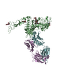
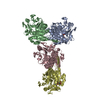
- PDB-6u3g: Best fitting antiparallel model for Volume 2 of truncated dimeric... -
+ Open data

- Basic information
| Entry | Database: PDB / ID: 6u3g | ||||||
|---|---|---|---|---|---|---|---|
| Title | Best fitting antiparallel model for Volume 2 of truncated dimeric Cytohesin-3 (Grp1; amino acids 14-399) | ||||||
 Components Components | Cytohesin-3 | ||||||
 Keywords Keywords | ENDOCYTOSIS / Arf GEF / Phosphoinositide binding / Sec7 Domain / PH Domain | ||||||
| Function / homology |  Function and homology information Function and homology informationestablishment of epithelial cell polarity / Golgi vesicle transport / Intra-Golgi traffic / regulation of ARF protein signal transduction / phosphatidylinositol-3,4,5-trisphosphate binding / bicellular tight junction / ruffle / positive regulation of cell adhesion / guanyl-nucleotide exchange factor activity / adherens junction ...establishment of epithelial cell polarity / Golgi vesicle transport / Intra-Golgi traffic / regulation of ARF protein signal transduction / phosphatidylinositol-3,4,5-trisphosphate binding / bicellular tight junction / ruffle / positive regulation of cell adhesion / guanyl-nucleotide exchange factor activity / adherens junction / Golgi membrane / nucleoplasm / plasma membrane / cytosol Similarity search - Function | ||||||
| Biological species |  Homo sapiens (human) Homo sapiens (human) | ||||||
| Method | ELECTRON MICROSCOPY / single particle reconstruction / negative staining / Resolution: 53 Å | ||||||
 Authors Authors | Das, S. / Lambright, D.G. | ||||||
| Funding support |  United States, 1items United States, 1items
| ||||||
 Citation Citation | Journal: Structure / Year: 2019 Title: Structural Organization and Dynamics of Homodimeric Cytohesin Family Arf GTPase Exchange Factors in Solution and on Membranes. Authors: Sanchaita Das / Andrew W Malaby / Agata Nawrotek / Wenhua Zhang / Mahel Zeghouf / Sarah Maslen / Mark Skehel / Srinivas Chakravarthy / Thomas C Irving / Osman Bilsel / Jacqueline Cherfils / ...Authors: Sanchaita Das / Andrew W Malaby / Agata Nawrotek / Wenhua Zhang / Mahel Zeghouf / Sarah Maslen / Mark Skehel / Srinivas Chakravarthy / Thomas C Irving / Osman Bilsel / Jacqueline Cherfils / David G Lambright /   Abstract: Membrane dynamic processes require Arf GTPase activation by guanine nucleotide exchange factors (GEFs) with a Sec7 domain. Cytohesin family Arf GEFs function in signaling and cell migration through ...Membrane dynamic processes require Arf GTPase activation by guanine nucleotide exchange factors (GEFs) with a Sec7 domain. Cytohesin family Arf GEFs function in signaling and cell migration through Arf GTPase activation on the plasma membrane and endosomes. In this study, the structural organization of two cytohesins (Grp1 and ARNO) was investigated in solution by size exclusion-small angle X-ray scattering and negative stain-electron microscopy and on membranes by dynamic light scattering, hydrogen-deuterium exchange-mass spectrometry and guanosine diphosphate (GDP)/guanosine triphosphate (GTP) exchange assays. The results suggest that cytohesins form elongated dimers with a central coiled coil and membrane-binding pleckstrin-homology (PH) domains at opposite ends. The dimers display significant conformational heterogeneity, with a preference for compact to intermediate conformations. Phosphoinositide-dependent membrane recruitment is mediated by one PH domain at a time and alters the conformational dynamics to prime allosteric activation by Arf-GTP. A structural model for membrane targeting and allosteric activation of full-length cytohesin dimers is discussed. | ||||||
| History |
|
- Structure visualization
| Movie |
Movie viewer |
|---|---|
| Structure viewer | Molecule: MolmilJmol/JSmol |
- Downloads & links
-Download
| PDBx/mmCIF format | 6u3g.cif.gz | 151.1 KB | Display | PDBx/mmCIF format |
|---|---|---|---|---|
| PDB format | pdb6u3g.ent.gz | 115.1 KB | Display | PDB format |
| PDBx/mmJSON format | 6u3g.json.gz | Tree view | PDBx/mmJSON format | |
| Others |  Other downloads Other downloads |
-Validation report
| Arichive directory | https://data.pdbj.org/pub/pdb/validation_reports/u3/6u3gftp://data.pdbj.org/pub/pdb/validation_reports/u3/6u3g | HTTPS FTP |
|---|
-Related structure data
| Related structure data |  20629MC  6u3eC M: map data used to model this data C: citing same article ( |
|---|---|
| Similar structure data | |
| Other databases |
|


-Links
 PDBj
PDBj
- Assembly
| Deposited unit | 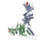
|
|---|---|
| 1 |
|
-Components
| #1: Protein | Mass: 46501.766 Da / Num. of mol.: 2 / Fragment: UNP residues 13-400 Source method: isolated from a genetically manipulated source Source: (gene. exp.) Homo sapiens (human) / Gene: CYTH3, ARNO3, GRP1, PSCD3 / Production host:  #2: Chemical | 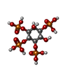  Mass: 500.075 Da / Num. of mol.: 2 / Source method: obtained synthetically / Formula: C6H16O18P4 Mass: 500.075 Da / Num. of mol.: 2 / Source method: obtained synthetically / Formula: C6H16O18P4Has ligand of interest | N | |
|---|
-Experimental details
-Experiment
| Experiment | Method: ELECTRON MICROSCOPY |
|---|---|
| EM experiment | Aggregation state: PARTICLE / 3D reconstruction method: single particle reconstruction |
- Sample preparation
| Component | Name: Truncated homodimer of Cytohesin-3 (Grp1, amino acids 14-399) with Inositol 1,3,4,5-tetrakis phosphate (IP4) Type: COMPLEX / Entity ID: #1 / Source: RECOMBINANT | |||||||||||||||
|---|---|---|---|---|---|---|---|---|---|---|---|---|---|---|---|---|
| Molecular weight | Experimental value: NO | |||||||||||||||
| Source (natural) | Organism: Homo sapiens (human) | |||||||||||||||
| Source (recombinant) | Organism: | |||||||||||||||
| Buffer solution | pH: 8 Details: Peak fractions after gel filtration were immediately diluted, applied to freshly glow discharged grids, and stained with uranyl formate. | |||||||||||||||
| Buffer component |
| |||||||||||||||
| Specimen | Embedding applied: NO / Shadowing applied: NO / Staining applied: YES / Vitrification applied: NO Details: The sample is a uniform homodimer with significant conformational flexibility. | |||||||||||||||
| EM staining | Type: NEGATIVE / Details: Stained with 0.75% (w/v) uranyl formate / Material: Uranyl Formate | |||||||||||||||
| Specimen support | Grid material: COPPER / Grid mesh size: 400 divisions/in. |
- Electron microscopy imaging
| Microscopy | Model: FEI/PHILIPS CM120T Details: specimen holder: FISCHIONE INSTRUMENTS DUAL AXIS TOMOGRAPHY HOLDER |
|---|---|
| Electron gun | Electron source:  FIELD EMISSION GUN / Accelerating voltage: 120 kV / Illumination mode: FLOOD BEAM FIELD EMISSION GUN / Accelerating voltage: 120 kV / Illumination mode: FLOOD BEAM |
| Electron lens | Mode: BRIGHT FIELD / Nominal magnification: 28000 X / Nominal defocus max: -3200 nm / Nominal defocus min: -1200 nm |
| Image recording | Electron dose: 30 e/Å2 / Film or detector model: TVIPS TEMCAM-F224 (2k x 2k) / Num. of grids imaged: 1 / Num. of real images: 500 |
| Image scans | Width: 2048 / Height: 2048 |
- Processing
| EM software |
| |||||||||||||||||||||||||||||||||||
|---|---|---|---|---|---|---|---|---|---|---|---|---|---|---|---|---|---|---|---|---|---|---|---|---|---|---|---|---|---|---|---|---|---|---|---|---|
| Image processing | Details: Images were processed with EMAN2 after X-ray removal with IMOD | |||||||||||||||||||||||||||||||||||
| CTF correction | Type: PHASE FLIPPING AND AMPLITUDE CORRECTION | |||||||||||||||||||||||||||||||||||
| Particle selection | Num. of particles selected: 6504 / Details: Particles were manually picked. | |||||||||||||||||||||||||||||||||||
| Symmetry | Point symmetry: C1 (asymmetric) | |||||||||||||||||||||||||||||||||||
| 3D reconstruction | Resolution: 53 Å / Resolution method: FSC 0.5 CUT-OFF / Num. of particles: 1863 / Algorithm: BACK PROJECTION / Num. of class averages: 15 / Symmetry type: POINT | |||||||||||||||||||||||||||||||||||
| Atomic model building | Protocol: RIGID BODY FIT / Space: REAL / Target criteria: Correlation coefficient Details: The model with the best correlation coefficient was selected by ADP_EM from a pool of 10000 models generated by RRT_SAMPLE using rigid bodies derived from 2R09 (autoinhibited core, amino ...Details: The model with the best correlation coefficient was selected by ADP_EM from a pool of 10000 models generated by RRT_SAMPLE using rigid bodies derived from 2R09 (autoinhibited core, amino acids 63-399) and a canonical antiparallel coiled coil (amino acids 18-53) built with CCBuilder. Flexible regions with reasonable geometry were modeled with MODELLER. | |||||||||||||||||||||||||||||||||||
| Atomic model building | PDB-ID: 2R09 Pdb chain-ID: B / Accession code: 2R09 / Pdb chain residue range: 63-399 / Source name: PDB / Type: experimental model |

