 Movie
Movie Controller

[English] 日本語
 Yorodumi
- PDB-6u1u: Human Angiopoietin-Like 4 C-Terminal Domain (cANGPTL4) with Palmi... -
+ Open data

- Basic information
| Entry | Database: PDB / ID: 6u1u | ||||||
|---|---|---|---|---|---|---|---|
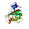
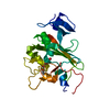
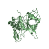
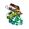
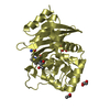
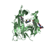
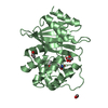
| Title | Human Angiopoietin-Like 4 C-Terminal Domain (cANGPTL4) with Palmitic Acid | ||||||
 Components Components | Angiopoietin-related protein 4 | ||||||
 Keywords Keywords | SIGNALING PROTEIN / Fibrinogen-like domain / Angiogenesis / Cancer cells / Metastasis | ||||||
| Function / homology |  Function and homology information Function and homology informationregulation of chylomicron remodeling / lipase inhibitor activity / lipase binding / Assembly of active LPL and LIPC lipase complexes / Regulation of CDH11 function / negative regulation of very-low-density lipoprotein particle remodeling / negative regulation of fatty acid biosynthetic process / endothelial cell apoptotic process / triglyceride homeostasis / protein unfolding ...regulation of chylomicron remodeling / lipase inhibitor activity / lipase binding / Assembly of active LPL and LIPC lipase complexes / Regulation of CDH11 function / negative regulation of very-low-density lipoprotein particle remodeling / negative regulation of fatty acid biosynthetic process / endothelial cell apoptotic process / triglyceride homeostasis / protein unfolding / negative regulation of endothelial cell apoptotic process / enzyme inhibitor activity / lipid metabolic process / PPARA activates gene expression / Transcriptional regulation of white adipocyte differentiation / positive regulation of angiogenesis / blood coagulation / MLL4 and MLL3 complexes regulate expression of PPARG target genes in adipogenesis and hepatic steatosis / angiogenesis / blood microparticle / response to hypoxia / negative regulation of apoptotic process / : / extracellular region / identical protein binding Similarity search - Function | ||||||
| Biological species |  Homo sapiens (human) Homo sapiens (human) | ||||||
| Method |  X-RAY DIFFRACTION / SYNCHROTRON / MOLECULAR REPLACEMENT / Resolution: 1.75 Å X-RAY DIFFRACTION / SYNCHROTRON / MOLECULAR REPLACEMENT / Resolution: 1.75 Å | ||||||
 Authors Authors | Tarver, C.L. / Yuan, Q. / Singhal, A.J. / Ramaker, R. / Cooper, S. / Pusey, M.L. | ||||||
| Funding support |  United States, 1items United States, 1items
| ||||||
 Citation Citation | Journal: To Be Published Title: Crystal Structures of Angiopoietin-Like 4 C-Terminal Domain (cANGPTL4) Reveal a Binding Pocket with Multiple Ligands Authors: Tarver, C.L. / Yuan, Q. / Singhal, A.J. / Ramaker, R. / Cooper, S. / Pusey, M.L. | ||||||
| History |
|
- Structure visualization
| Structure viewer | Molecule: MolmilJmol/JSmol |
|---|
- Downloads & links
-Download
| PDBx/mmCIF format | 6u1u.cif.gz | 62.4 KB | Display | PDBx/mmCIF format |
|---|---|---|---|---|
| PDB format | pdb6u1u.ent.gz | 42.6 KB | Display | PDB format |
| PDBx/mmJSON format | 6u1u.json.gz | Tree view | PDBx/mmJSON format | |
| Others |  Other downloads Other downloads |
-Validation report
| Arichive directory | https://data.pdbj.org/pub/pdb/validation_reports/u1/6u1uftp://data.pdbj.org/pub/pdb/validation_reports/u1/6u1u | HTTPS FTP |
|---|
-Related structure data
| Related structure data |  6u0aC  6eubS S: Starting model for refinement C: citing same article ( |
|---|---|
| Similar structure data |


-Links
 PDBj
PDBj


- Assembly
| Deposited unit | 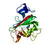
| ||||||||||||
|---|---|---|---|---|---|---|---|---|---|---|---|---|---|
| 1 |
| ||||||||||||
| Unit cell |
|
-Components
| #1: Protein | Mass: 24458.404 Da / Num. of mol.: 1 Source method: isolated from a genetically manipulated source Source: (gene. exp.) Homo sapiens (human)Gene: ANGPTL4, ARP4, HFARP, PGAR, PP1158, PSEC0166, UNQ171/PRO197 Plasmid: pET3a / Production host:  |
|---|---|
| #2: Chemical | ChemComp-PLM /   Mass: 256.424 Da / Num. of mol.: 1 / Source method: obtained synthetically / Formula: C16H32O2 / Feature type: SUBJECT OF INVESTIGATION Mass: 256.424 Da / Num. of mol.: 1 / Source method: obtained synthetically / Formula: C16H32O2 / Feature type: SUBJECT OF INVESTIGATION |
| #3: Water | ChemComp-HOH /  Mass: 18.015 Da / Num. of mol.: 122 / Source method: isolated from a natural source / Formula: H2O Mass: 18.015 Da / Num. of mol.: 122 / Source method: isolated from a natural source / Formula: H2O |
| Has ligand of interest | Y |
| Has protein modification | Y |
-Experimental details
-Experiment
| Experiment | Method: X-RAY DIFFRACTION / Number of used crystals: 1 |
|---|
- Sample preparation
| Crystal | Density Matthews: 3.62 Å3/Da / Density % sol: 66.04 % |
|---|---|
| Crystal grow | Temperature: 295 K / Method: vapor diffusion, sitting drop Details: 0.1 M HEPES 7.5 0.1 M Sodium chloride 1.6 M Ammonium sulfate Palmitic Acid PH range: 7.5-8.5 |
-Data collection
| Diffraction | Mean temperature: 100 K / Serial crystal experiment: N |
|---|---|
| Diffraction source | Source: SYNCHROTRON / Site: APS / Beamline: 19-ID / Wavelength: 0.988 Å |
| Detector | Type: DECTRIS PILATUS3 S 6M / Detector: PIXEL / Date: Jun 29, 2019 |
| Radiation | Protocol: SINGLE WAVELENGTH / Monochromatic (M) / Laue (L): M / Scattering type: x-ray |
| Radiation wavelength | Wavelength: 0.988 Å / Relative weight: 1 |
| Reflection | Resolution: 1.7→42.7 Å / Num. obs: 34756 / % possible obs: 93.12 % / Redundancy: 6.5 % / Biso Wilson estimate: 24.34 Å2 / CC1/2: 0.998 / Rmerge(I) obs: 0.054 / Rpim(I) all: 0.023 / Rrim(I) all: 0.059 / Net I/σ(I): 15.7 |
| Reflection shell | Resolution: 1.75→1.78 Å / Rmerge(I) obs: 0.62 / Num. unique obs: 1727 / CC1/2: 0.849 / Rpim(I) all: 0.264 / Rrim(I) all: 0.675 |
- Processing
| Software |
| |||||||||||||||||||||||||||||||||||||||||||||||||||||||||||||||||||||||||||||||||||||||||||||||||||||||||
|---|---|---|---|---|---|---|---|---|---|---|---|---|---|---|---|---|---|---|---|---|---|---|---|---|---|---|---|---|---|---|---|---|---|---|---|---|---|---|---|---|---|---|---|---|---|---|---|---|---|---|---|---|---|---|---|---|---|---|---|---|---|---|---|---|---|---|---|---|---|---|---|---|---|---|---|---|---|---|---|---|---|---|---|---|---|---|---|---|---|---|---|---|---|---|---|---|---|---|---|---|---|---|---|---|---|---|
| Refinement | Method to determine structure: MOLECULAR REPLACEMENT Starting model: 6EUB Resolution: 1.75→42.7 Å / SU ML: 0.1777 / Cross valid method: FREE R-VALUE / σ(F): 0 / Phase error: 19.3847
| |||||||||||||||||||||||||||||||||||||||||||||||||||||||||||||||||||||||||||||||||||||||||||||||||||||||||
| Solvent computation | Shrinkage radii: 0.9 Å / VDW probe radii: 1.11 Å | |||||||||||||||||||||||||||||||||||||||||||||||||||||||||||||||||||||||||||||||||||||||||||||||||||||||||
| Displacement parameters | Biso mean: 29.12 Å2 | |||||||||||||||||||||||||||||||||||||||||||||||||||||||||||||||||||||||||||||||||||||||||||||||||||||||||
| Refinement step | Cycle: LAST / Resolution: 1.75→42.7 Å
| |||||||||||||||||||||||||||||||||||||||||||||||||||||||||||||||||||||||||||||||||||||||||||||||||||||||||
| Refine LS restraints |
| |||||||||||||||||||||||||||||||||||||||||||||||||||||||||||||||||||||||||||||||||||||||||||||||||||||||||
| LS refinement shell |
|
