 Movie
Movie Controller

[English] 日本語
 Yorodumi
- PDB-6twl: Apo structure of the Ectoine utilization protein EutE (DoeB) from... -
+ Open data

- Basic information
| Entry | Database: PDB / ID: 6twl | ||||||
|---|---|---|---|---|---|---|---|
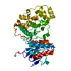
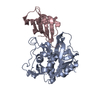
| Title | Apo structure of the Ectoine utilization protein EutE (DoeB) from Ruegeria pomeroyi | ||||||
 Components Components | N-acetyl-L-2,4-diaminobutyric acid deacetylase | ||||||
 Keywords Keywords | HYDROLASE / Pita bread / ectoine degradation | ||||||
| Function / homology |  Function and homology information Function and homology informationhydrolase activity, acting on carbon-nitrogen (but not peptide) bonds, in linear amides / hydrolase activity, acting on ester bonds / metal ion binding Similarity search - Function | ||||||
| Biological species |  Ruegeria pomeroyi (bacteria) Ruegeria pomeroyi (bacteria) | ||||||
| Method |  X-RAY DIFFRACTION / SYNCHROTRON / MOLECULAR REPLACEMENT / Resolution: 2 Å X-RAY DIFFRACTION / SYNCHROTRON / MOLECULAR REPLACEMENT / Resolution: 2 Å | ||||||
 Authors Authors | Mais, C.-N. / Altegoer, F. / Bange, G. | ||||||
 Citation Citation | Journal: J.Biol.Chem. / Year: 2020 Title: Degradation of the microbial stress protectants and chemical chaperones ectoine and hydroxyectoine by a bacterial hydrolase-deacetylase complex. Authors: Mais, C.N. / Hermann, L. / Altegoer, F. / Seubert, A. / Richter, A.A. / Wernersbach, I. / Czech, L. / Bremer, E. / Bange, G. | ||||||
| History |
|
- Structure visualization
| Structure viewer | Molecule: MolmilJmol/JSmol |
|---|
- Downloads & links
-Download
| PDBx/mmCIF format | 6twl.cif.gz | 154.8 KB | Display | PDBx/mmCIF format |
|---|---|---|---|---|
| PDB format | pdb6twl.ent.gz | 103.5 KB | Display | PDB format |
| PDBx/mmJSON format | 6twl.json.gz | Tree view | PDBx/mmJSON format | |
| Others |  Other downloads Other downloads |
-Validation report
| Arichive directory | https://data.pdbj.org/pub/pdb/validation_reports/tw/6twlftp://data.pdbj.org/pub/pdb/validation_reports/tw/6twl | HTTPS FTP |
|---|
-Related structure data
| Related structure data |  6twjC  6twkC  6twmC  6yo9C  3cdxS S: Starting model for refinement C: citing same article ( |
|---|---|
| Similar structure data |


-Links
 PDBj
PDBj- Assembly
| Deposited unit | 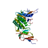
| ||||||||||||
|---|---|---|---|---|---|---|---|---|---|---|---|---|---|
| 1 |
| ||||||||||||
| 2 | x 6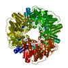
| ||||||||||||
| Unit cell |
|
-Components
| #1: Protein | Mass: 35146.949 Da / Num. of mol.: 1 Source method: isolated from a genetically manipulated source Source: (gene. exp.) Ruegeria pomeroyi (strain ATCC 700808 / DSM 15171 / DSS-3) (bacteria)Gene: doeB, SPO1139 / Production host: |
|---|---|
| #2: Water | ChemComp-HOH /  Mass: 18.015 Da / Num. of mol.: 14 / Source method: isolated from a natural source / Formula: H2O Mass: 18.015 Da / Num. of mol.: 14 / Source method: isolated from a natural source / Formula: H2O |
-Experimental details
-Experiment
| Experiment | Method: X-RAY DIFFRACTION / Number of used crystals: 1 |
|---|
- Sample preparation
| Crystal | Density Matthews: 2.72 Å3/Da / Density % sol: 54.75 % |
|---|---|
| Crystal grow | Temperature: 293 K / Method: vapor diffusion, sitting drop / pH: 9 / Details: 0.1 M Bicine pH 9.0, 20% (wt/vol) PEG 6000 |
-Data collection
| Diffraction | Mean temperature: 100 K / Serial crystal experiment: N |
|---|---|
| Diffraction source | Source: SYNCHROTRON / Site: ESRF  / Beamline: ID23-1 / Wavelength: 0.97242 Å / Beamline: ID23-1 / Wavelength: 0.97242 Å |
| Detector | Type: DECTRIS PILATUS 6M / Detector: PIXEL / Date: Dec 6, 2018 |
| Radiation | Protocol: SINGLE WAVELENGTH / Monochromatic (M) / Laue (L): M / Scattering type: x-ray |
| Radiation wavelength | Wavelength: 0.97242 Å / Relative weight: 1 |
| Reflection | Resolution: 1.99→46.54 Å / Num. obs: 27066 / % possible obs: 99.48 % / Redundancy: 21 % / Biso Wilson estimate: 61.44 Å2 / CC1/2: 1 / Rmerge(I) obs: 0.0626 / Net I/σ(I): 23 |
| Reflection shell | Resolution: 1.99→2.06 Å / Mean I/σ(I) obs: 0.6 / Num. unique obs: 2612 / CC1/2: 0.551 |
- Processing
| Software |
| |||||||||||||||||||||||||||||||||||||||||||||||||||||||||||||||||||||||||||||||||||||||||||||||||||||||||||||||||||||||||||||
|---|---|---|---|---|---|---|---|---|---|---|---|---|---|---|---|---|---|---|---|---|---|---|---|---|---|---|---|---|---|---|---|---|---|---|---|---|---|---|---|---|---|---|---|---|---|---|---|---|---|---|---|---|---|---|---|---|---|---|---|---|---|---|---|---|---|---|---|---|---|---|---|---|---|---|---|---|---|---|---|---|---|---|---|---|---|---|---|---|---|---|---|---|---|---|---|---|---|---|---|---|---|---|---|---|---|---|---|---|---|---|---|---|---|---|---|---|---|---|---|---|---|---|---|---|---|---|
| Refinement | Method to determine structure: MOLECULAR REPLACEMENT Starting model: 3CDX Resolution: 2→46.54 Å / SU ML: 0.3627 / Cross valid method: FREE R-VALUE / σ(F): 1.33 / Phase error: 41.4651 Stereochemistry target values: GeoStd + Monomer Library + CDL v1.2
| |||||||||||||||||||||||||||||||||||||||||||||||||||||||||||||||||||||||||||||||||||||||||||||||||||||||||||||||||||||||||||||
| Solvent computation | Shrinkage radii: 0.9 Å / VDW probe radii: 1.11 Å / Solvent model: FLAT BULK SOLVENT MODEL | |||||||||||||||||||||||||||||||||||||||||||||||||||||||||||||||||||||||||||||||||||||||||||||||||||||||||||||||||||||||||||||
| Displacement parameters | Biso mean: 80.66 Å2 | |||||||||||||||||||||||||||||||||||||||||||||||||||||||||||||||||||||||||||||||||||||||||||||||||||||||||||||||||||||||||||||
| Refinement step | Cycle: LAST / Resolution: 2→46.54 Å
| |||||||||||||||||||||||||||||||||||||||||||||||||||||||||||||||||||||||||||||||||||||||||||||||||||||||||||||||||||||||||||||
| Refine LS restraints |
| |||||||||||||||||||||||||||||||||||||||||||||||||||||||||||||||||||||||||||||||||||||||||||||||||||||||||||||||||||||||||||||
| LS refinement shell |
| |||||||||||||||||||||||||||||||||||||||||||||||||||||||||||||||||||||||||||||||||||||||||||||||||||||||||||||||||||||||||||||
| Refinement TLS params. | Method: refined / Refine-ID: X-RAY DIFFRACTION
| |||||||||||||||||||||||||||||||||||||||||||||||||||||||||||||||||||||||||||||||||||||||||||||||||||||||||||||||||||||||||||||
| Refinement TLS group |
|
