 Movie
Movie Controller

[English] 日本語
 Yorodumi
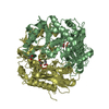
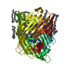
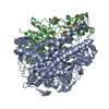
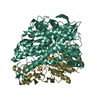
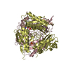
- PDB-6twk: Substrate bound structure of the Ectoine utilization protein EutD... -
+ Open data

- Basic information
| Entry | Database: PDB / ID: 6twk | ||||||
|---|---|---|---|---|---|---|---|
| Title | Substrate bound structure of the Ectoine utilization protein EutD (DoeA) from Halomonas elongata | ||||||
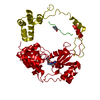
 Components Components | Ectoine hydrolase DoeA | ||||||
 Keywords Keywords | HYDROLASE / Pita bread / ectoine degradation | ||||||
| Function / homology |  Function and homology information Function and homology informationectoine hydrolase / ectoine catabolic process / hydrolase activity, acting on carbon-nitrogen (but not peptide) bonds, in cyclic amides / Hydrolases; Acting on peptide bonds (peptidases) / hydrolase activity / cytoplasm Similarity search - Function | ||||||
| Biological species |  Halomonas elongata (bacteria) Halomonas elongata (bacteria) | ||||||
| Method |  X-RAY DIFFRACTION / SYNCHROTRON / MOLECULAR REPLACEMENT / Resolution: 2.25 Å X-RAY DIFFRACTION / SYNCHROTRON / MOLECULAR REPLACEMENT / Resolution: 2.25 Å | ||||||
 Authors Authors | Mais, C.-N. / Altegoer, F. / Bange, G. | ||||||
 Citation Citation | Journal: J.Biol.Chem. / Year: 2020 Title: Degradation of the microbial stress protectants and chemical chaperones ectoine and hydroxyectoine by a bacterial hydrolase-deacetylase complex. Authors: Mais, C.N. / Hermann, L. / Altegoer, F. / Seubert, A. / Richter, A.A. / Wernersbach, I. / Czech, L. / Bremer, E. / Bange, G. | ||||||
| History |
|
- Structure visualization
| Structure viewer | Molecule: MolmilJmol/JSmol |
|---|
- Downloads & links
-Download
| PDBx/mmCIF format | 6twk.cif.gz | 182.6 KB | Display | PDBx/mmCIF format |
|---|---|---|---|---|
| PDB format | pdb6twk.ent.gz | 140.8 KB | Display | PDB format |
| PDBx/mmJSON format | 6twk.json.gz | Tree view | PDBx/mmJSON format | |
| Others |  Other downloads Other downloads |
-Validation report
| Arichive directory | https://data.pdbj.org/pub/pdb/validation_reports/tw/6twkftp://data.pdbj.org/pub/pdb/validation_reports/tw/6twk | HTTPS FTP |
|---|
-Related structure data
| Related structure data | 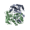 6twjSC  6twlC 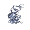 6twmC 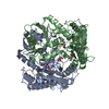 6yo9C S: Starting model for refinement C: citing same article ( |
|---|---|
| Similar structure data |


-Links
 PDBj
PDBj- Assembly
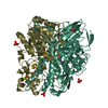
| Deposited unit | 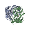
| ||||||||||||
|---|---|---|---|---|---|---|---|---|---|---|---|---|---|
| 1 |
| ||||||||||||
| Unit cell |
| ||||||||||||
| Components on special symmetry positions |
|
-Components
| #1: Protein | Mass: 44995.125 Da / Num. of mol.: 2 Source method: isolated from a genetically manipulated source Source: (gene. exp.) Halomonas elongata (bacteria) / Gene: doeA, A8U91_03446, DKQ62_09665 / Production host: References: UniProt: A0A1B8NWR1, UniProt: E1V7W1*PLUS, Hydrolases; Acting on peptide bonds (peptidases) #2: Chemical | ChemComp-P4B / ( | 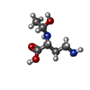  Mass: 162.187 Da / Num. of mol.: 1 / Source method: isolated from a natural source / Formula: C6H14N2O3 / Feature type: SUBJECT OF INVESTIGATION Mass: 162.187 Da / Num. of mol.: 1 / Source method: isolated from a natural source / Formula: C6H14N2O3 / Feature type: SUBJECT OF INVESTIGATION#3: Chemical | ChemComp-4CS / ( |   Mass: 142.156 Da / Num. of mol.: 1 / Source method: isolated from a natural source / Formula: C6H10N2O2 / Feature type: SUBJECT OF INVESTIGATION Mass: 142.156 Da / Num. of mol.: 1 / Source method: isolated from a natural source / Formula: C6H10N2O2 / Feature type: SUBJECT OF INVESTIGATION#4: Water | ChemComp-HOH / |  Mass: 18.015 Da / Num. of mol.: 523 / Source method: isolated from a natural source / Formula: H2O Mass: 18.015 Da / Num. of mol.: 523 / Source method: isolated from a natural source / Formula: H2OHas ligand of interest | Y | |
|---|
-Experimental details
-Experiment
| Experiment | Method: X-RAY DIFFRACTION / Number of used crystals: 1 |
|---|
- Sample preparation
| Crystal | Density Matthews: 4.17 Å3/Da / Density % sol: 70.5 % |
|---|---|
| Crystal grow | Temperature: 293 K / Method: vapor diffusion, sitting drop / Details: 0.2 M trisodium citrate, 20% (wt/vol) PEG 3350 / PH range: 6.5-7.5 |
-Data collection
| Diffraction | Mean temperature: 100 K / Serial crystal experiment: N |
|---|---|
| Diffraction source | Source: SYNCHROTRON / Site: ESRF  / Beamline: ID23-2 / Wavelength: 0.873127 Å / Beamline: ID23-2 / Wavelength: 0.873127 Å |
| Detector | Type: DECTRIS PILATUS 6M / Detector: PIXEL / Date: Sep 26, 2019 |
| Radiation | Protocol: SINGLE WAVELENGTH / Monochromatic (M) / Laue (L): M / Scattering type: x-ray |
| Radiation wavelength | Wavelength: 0.873127 Å / Relative weight: 1 |
| Reflection | Resolution: 2.2→48.42 Å / Num. obs: 78809 / % possible obs: 99.96 % / Redundancy: 8.9 % / Biso Wilson estimate: 34.55 Å2 / CC1/2: 0.997 / Rmerge(I) obs: 0.1474 / Net I/σ(I): 10.69 |
| Reflection shell | Resolution: 2.2→2.279 Å / Rmerge(I) obs: 1.466 / Mean I/σ(I) obs: 1.65 / Num. unique obs: 6599 / CC1/2: 0.583 |
- Processing
| Software |
| ||||||||||||||||||||||||
|---|---|---|---|---|---|---|---|---|---|---|---|---|---|---|---|---|---|---|---|---|---|---|---|---|---|
| Refinement | Method to determine structure: MOLECULAR REPLACEMENT Starting model: 6TWJ Resolution: 2.25→43.88 Å / Cross valid method: FREE R-VALUE
| ||||||||||||||||||||||||
| Displacement parameters | Biso mean: 38.52 Å2 | ||||||||||||||||||||||||
| Refinement step | Cycle: LAST / Resolution: 2.25→43.88 Å
| ||||||||||||||||||||||||
| Refine LS restraints |
|
