 Movie
Movie Controller

[English] 日本語
 Yorodumi
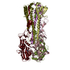
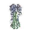
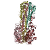
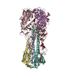
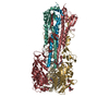
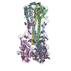
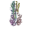
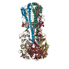
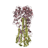
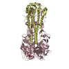
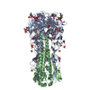
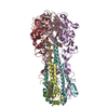
- PDB-6tvd: Crystal structure of the haemagglutinin from a H10N7 seal influen... -
+ Open data

- Basic information
| Entry | Database: PDB / ID: 6tvd | ||||||
|---|---|---|---|---|---|---|---|
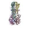
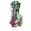
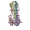
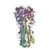
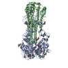
| Title | Crystal structure of the haemagglutinin from a H10N7 seal influenza virus isolated in Germany in complex with avian receptor analogue, 3'-SLN | ||||||
 Components Components | (Hemagglutinin ...) x 2 | ||||||
 Keywords Keywords | VIRAL PROTEIN / receptor binding / fusion of virus membrane with host plasma membrane | ||||||
| Function / homology |  Function and homology information Function and homology informationclathrin-dependent endocytosis of virus by host cell / host cell surface receptor binding / fusion of virus membrane with host plasma membrane / fusion of virus membrane with host endosome membrane / viral envelope / virion attachment to host cell / host cell plasma membrane / metal ion binding Similarity search - Function | ||||||
| Biological species |   Influenza A virus Influenza A virus | ||||||
| Method |  X-RAY DIFFRACTION / SYNCHROTRON / MOLECULAR REPLACEMENT / Resolution: 2.7 Å X-RAY DIFFRACTION / SYNCHROTRON / MOLECULAR REPLACEMENT / Resolution: 2.7 Å | ||||||
 Authors Authors | Zhang, J. / Xiong, X. / Purkiss, A. / Walker, P. / Gamblin, S. / Skehel, J.J. | ||||||
| Funding support |  United Kingdom, 1items United Kingdom, 1items
| ||||||
 Citation Citation | Journal: Cell Host Microbe / Year: 2020 Title: Hemagglutinin Traits Determine Transmission of Avian A/H10N7 Influenza Virus between Mammals. Authors: Herfst, S. / Zhang, J. / Richard, M. / McBride, R. / Lexmond, P. / Bestebroer, T.M. / Spronken, M.I.J. / de Meulder, D. / van den Brand, J.M. / Rosu, M.E. / Martin, S.R. / Gamblin, S.J. / ...Authors: Herfst, S. / Zhang, J. / Richard, M. / McBride, R. / Lexmond, P. / Bestebroer, T.M. / Spronken, M.I.J. / de Meulder, D. / van den Brand, J.M. / Rosu, M.E. / Martin, S.R. / Gamblin, S.J. / Xiong, X. / Peng, W. / Bodewes, R. / van der Vries, E. / Osterhaus, A.D.M.E. / Paulson, J.C. / Skehel, J.J. / Fouchier, R.A.M. | ||||||
| History |
|
- Structure visualization
| Structure viewer | Molecule: MolmilJmol/JSmol |
|---|
- Downloads & links
-Download
| PDBx/mmCIF format | 6tvd.cif.gz | 585.3 KB | Display | PDBx/mmCIF format |
|---|---|---|---|---|
| PDB format | pdb6tvd.ent.gz | 484.6 KB | Display | PDB format |
| PDBx/mmJSON format | 6tvd.json.gz | Tree view | PDBx/mmJSON format | |
| Others |  Other downloads Other downloads |
-Validation report
| Arichive directory | https://data.pdbj.org/pub/pdb/validation_reports/tv/6tvdftp://data.pdbj.org/pub/pdb/validation_reports/tv/6tvd | HTTPS FTP |
|---|
-Related structure data
| Related structure data | 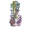 6tjwC 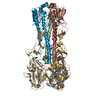 6tjyC 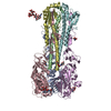 6tvaC 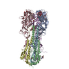 6tvbC 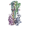 6tvcC 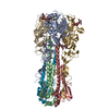 6tvfC 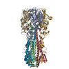 6tvrC 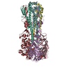 6tvsC  6tvtC 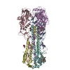 6twhC 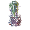 6twiC 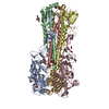 6twsC 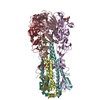 6twvC 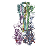 6txoC 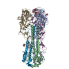 6ty1C 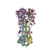 4d00S S: Starting model for refinement C: citing same article ( |
|---|---|
| Similar structure data |


-Links
 PDBj
PDBj


- Assembly
| Deposited unit | 
| ||||||||
|---|---|---|---|---|---|---|---|---|---|
| 1 | 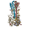
| ||||||||
| 2 | 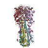
| ||||||||
| Unit cell |
|
-Components
-Hemagglutinin ... , 2 types, 12 molecules ACEGIKBDFHJL
| #1: Protein | Mass: 35499.078 Da / Num. of mol.: 6 Source method: isolated from a genetically manipulated source Source: (gene. exp.) Influenza A virus / Gene: HA / Cell line (production host): Sf9 / Production host: unidentified baculovirus / References: UniProt: A0A0A7HR51#2: Protein | Mass: 20313.389 Da / Num. of mol.: 6 Source method: isolated from a genetically manipulated source Source: (gene. exp.) Influenza A virus / Gene: HA / Cell line (production host): Sf9 / Production host: unidentified baculovirus / References: UniProt: A0A0A7HR51 |
|---|
-Sugars , 4 types, 20 molecules 
| #3: Polysaccharide | N-acetyl-alpha-neuraminic acid-(2-3)-beta-D-galactopyranose-(1-4)-2-acetamido-2-deoxy-beta-D-glucopyranose / 3'-sialyl-N-acetyllactosamine 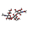  Source method: isolated from a genetically manipulated source Details: oligosaccharide / References: 3'-sialyl-N-acetyllactosamine #4: Polysaccharide | 2-acetamido-2-deoxy-beta-D-glucopyranose-(1-4)-2-acetamido-2-deoxy-beta-D-glucopyranose Source method: isolated from a genetically manipulated source #5: Polysaccharide | N-acetyl-alpha-neuraminic acid-(2-3)-beta-D-galactopyranose | Source method: isolated from a genetically manipulated source #6: Sugar | ChemComp-NAG /  Type: D-saccharide, beta linking / Mass: 221.208 Da / Num. of mol.: 9 Type: D-saccharide, beta linking / Mass: 221.208 Da / Num. of mol.: 9Source method: isolated from a genetically manipulated source Formula: C8H15NO6 |
|---|
-Non-polymers , 2 types, 158 molecules 

| #7: Chemical |  Mass: 40.078 Da / Num. of mol.: 3 / Source method: obtained synthetically / Formula: Ca Mass: 40.078 Da / Num. of mol.: 3 / Source method: obtained synthetically / Formula: Ca#8: Water | ChemComp-HOH / | Mass: 18.015 Da / Num. of mol.: 155 / Source method: isolated from a natural source / Formula: H2O |
|---|
-Details
| Has ligand of interest | N |
|---|---|
| Has protein modification | Y |
-Experimental details
-Experiment
| Experiment | Method: X-RAY DIFFRACTION / Number of used crystals: 1 |
|---|
- Sample preparation
| Crystal | Density Matthews: 3.5 Å3/Da / Density % sol: 65 % |
|---|---|
| Crystal grow | Temperature: 298 K / Method: vapor diffusion, sitting drop / Details: 2%-4% PEG6000, 0.1M HEPES pH7.0 |
-Data collection
| Diffraction | Mean temperature: 100 K / Serial crystal experiment: N |
|---|---|
| Diffraction source | Source: SYNCHROTRON / Site: Diamond / Beamline: I02 / Wavelength: 0.9795 Å |
| Detector | Type: DECTRIS PILATUS 6M-F / Detector: PIXEL / Date: Nov 27, 2015 |
| Radiation | Protocol: SINGLE WAVELENGTH / Monochromatic (M) / Laue (L): M / Scattering type: x-ray |
| Radiation wavelength | Wavelength: 0.9795 Å / Relative weight: 1 |
| Reflection | Resolution: 2.7→68.55 Å / Num. obs: 123642 / % possible obs: 99.1 % / Redundancy: 3 % / CC1/2: 0.994 / Net I/σ(I): 10.4 |
| Reflection shell | Resolution: 2.7→2.75 Å / Num. unique obs: 6123 / CC1/2: 0.503 |
- Processing
| Software |
| |||||||||||||||||||||||||||||||||||||||||||||||||||||||||||||||||||||||||||
|---|---|---|---|---|---|---|---|---|---|---|---|---|---|---|---|---|---|---|---|---|---|---|---|---|---|---|---|---|---|---|---|---|---|---|---|---|---|---|---|---|---|---|---|---|---|---|---|---|---|---|---|---|---|---|---|---|---|---|---|---|---|---|---|---|---|---|---|---|---|---|---|---|---|---|---|---|
| Refinement | Method to determine structure: MOLECULAR REPLACEMENT Starting model: 4D00 Resolution: 2.7→68.55 Å / Cor.coef. Fo:Fc: 0.935 / Cor.coef. Fo:Fc free: 0.911 / Occupancy max: 1 / Occupancy min: 0.03 / Cross valid method: THROUGHOUT / σ(F): 0 / ESU R: 0.677 / ESU R Free: 0.386 / Stereochemistry target values: MAXIMUM LIKELIHOOD Details: HYDROGENS HAVE BEEN ADDED IN THE RIDING POSITIONS U VALUES : REFINED INDIVIDUALLY
| |||||||||||||||||||||||||||||||||||||||||||||||||||||||||||||||||||||||||||
| Solvent computation | Ion probe radii: 0.8 Å / Shrinkage radii: 0.8 Å / VDW probe radii: 1.2 Å / Solvent model: MASK | |||||||||||||||||||||||||||||||||||||||||||||||||||||||||||||||||||||||||||
| Displacement parameters | Biso max: 244.89 Å2 / Biso mean: 105.153 Å2 / Biso min: 43.46 Å2
| |||||||||||||||||||||||||||||||||||||||||||||||||||||||||||||||||||||||||||
| Refinement step | Cycle: LAST / Resolution: 2.7→68.55 Å
| |||||||||||||||||||||||||||||||||||||||||||||||||||||||||||||||||||||||||||
| Refine LS restraints |
| |||||||||||||||||||||||||||||||||||||||||||||||||||||||||||||||||||||||||||
| LS refinement shell | Resolution: 2.7→2.77 Å / Total num. of bins used: 20
|
