 Movie
Movie Controller

[English] 日本語
 Yorodumi
- PDB-6tr4: Ruminococcus gnavus GH29 fucosidase E1_10125 D221A mutant in comp... -
+ Open data

- Basic information
| Entry | Database: PDB / ID: 6tr4 | |||||||||
|---|---|---|---|---|---|---|---|---|---|---|
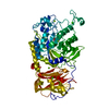
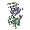
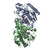
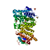
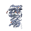
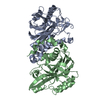
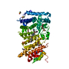
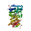
| Title | Ruminococcus gnavus GH29 fucosidase E1_10125 D221A mutant in complex with fucose | |||||||||
 Components Components | F5/8 type C domain-containing protein | |||||||||
 Keywords Keywords | HYDROLASE / fucosidase / fucose / ruminococcus gnavus / GH29 | |||||||||
| Function / homology |  Function and homology information Function and homology informationalpha-L-fucosidase / alpha-L-fucosidase activity / fucose metabolic process / glycoside catabolic process / lysosome / metal ion binding Similarity search - Function | |||||||||
| Biological species |  [Ruminococcus] gnavus E1 (bacteria) [Ruminococcus] gnavus E1 (bacteria) | |||||||||
| Method |  X-RAY DIFFRACTION / SYNCHROTRON / MOLECULAR REPLACEMENT / Resolution: 1.45 Å X-RAY DIFFRACTION / SYNCHROTRON / MOLECULAR REPLACEMENT / Resolution: 1.45 Å | |||||||||
 Authors Authors | Owen, C.D. / Wu, H. / Crost, E. / Colvile, A. / Juge, N. / Walsh, M.A. | |||||||||
| Funding support |  United Kingdom, 2items United Kingdom, 2items
| |||||||||
 Citation Citation | Journal: Cell.Mol.Life Sci. / Year: 2021 Title: Fucosidases from the human gut symbiont Ruminococcus gnavus. Authors: Wu, H. / Rebello, O. / Crost, E.H. / Owen, C.D. / Walpole, S. / Bennati-Granier, C. / Ndeh, D. / Monaco, S. / Hicks, T. / Colvile, A. / Urbanowicz, P.A. / Walsh, M.A. / Angulo, J. / Spencer, D.I.R. / Juge, N. | |||||||||
| History |
|
- Structure visualization
| Structure viewer | Molecule: MolmilJmol/JSmol |
|---|
- Downloads & links
-Download
| PDBx/mmCIF format | 6tr4.cif.gz | 432.5 KB | Display | PDBx/mmCIF format |
|---|---|---|---|---|
| PDB format | pdb6tr4.ent.gz | 348.4 KB | Display | PDB format |
| PDBx/mmJSON format | 6tr4.json.gz | Tree view | PDBx/mmJSON format | |
| Others |  Other downloads Other downloads |
-Validation report
| Arichive directory | https://data.pdbj.org/pub/pdb/validation_reports/tr/6tr4ftp://data.pdbj.org/pub/pdb/validation_reports/tr/6tr4 | HTTPS FTP |
|---|
-Related structure data
| Related structure data |  6tr3C  4oueS S: Starting model for refinement C: citing same article ( |
|---|---|
| Similar structure data |


-Links
 PDBj
PDBj


- Assembly
| Deposited unit | 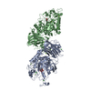
| ||||||||||||||||||
|---|---|---|---|---|---|---|---|---|---|---|---|---|---|---|---|---|---|---|---|
| 1 | 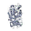
| ||||||||||||||||||
| 2 | 
| ||||||||||||||||||
| Unit cell |
| ||||||||||||||||||
| Noncrystallographic symmetry (NCS) | NCS domain:
NCS domain segments: Component-ID: _ / Ens-ID: 1 / Beg auth comp-ID: ASN / Beg label comp-ID: ASN / End auth comp-ID: PRO / End label comp-ID: PRO / Refine code: _ / Auth seq-ID: 7 - 512 / Label seq-ID: 7 - 512
|
-Components
-Protein , 1 types, 2 molecules AB
| #1: Protein | Mass: 61664.961 Da / Num. of mol.: 2 Source method: isolated from a genetically manipulated source Source: (gene. exp.) [Ruminococcus] gnavus E1 (bacteria) / Gene: CDL26_02305 / Production host: |
|---|
-Sugars , 2 types, 3 molecules 

| #2: Sugar | ChemComp-FUL /  Type: L-saccharide, beta linking / Mass: 164.156 Da / Num. of mol.: 1 Type: L-saccharide, beta linking / Mass: 164.156 Da / Num. of mol.: 1Source method: isolated from a genetically manipulated source Formula: C6H12O5 / Feature type: SUBJECT OF INVESTIGATION |
|---|---|
| #3: Sugar |  Type: L-saccharide, alpha linking / Mass: 164.156 Da / Num. of mol.: 2 Type: L-saccharide, alpha linking / Mass: 164.156 Da / Num. of mol.: 2Source method: isolated from a genetically manipulated source Formula: C6H12O5 / Feature type: SUBJECT OF INVESTIGATION |
-Non-polymers , 4 types, 1522 molecules 


| #4: Chemical | ChemComp-MG /  Mass: 24.305 Da / Num. of mol.: 5 / Source method: obtained synthetically / Formula: Mg Mass: 24.305 Da / Num. of mol.: 5 / Source method: obtained synthetically / Formula: Mg#5: Chemical | ChemComp-CA /  Mass: 40.078 Da / Num. of mol.: 6 / Source method: obtained synthetically / Formula: Ca Mass: 40.078 Da / Num. of mol.: 6 / Source method: obtained synthetically / Formula: Ca#6: Chemical | ChemComp-CL /  Mass: 35.453 Da / Num. of mol.: 4 / Source method: obtained synthetically / Formula: Cl Mass: 35.453 Da / Num. of mol.: 4 / Source method: obtained synthetically / Formula: Cl#7: Water | ChemComp-HOH / | Mass: 18.015 Da / Num. of mol.: 1507 / Source method: isolated from a natural source / Formula: H2O |
|---|
-Details
| Has ligand of interest | Y |
|---|
-Experimental details
-Experiment
| Experiment | Method: X-RAY DIFFRACTION / Number of used crystals: 1 |
|---|
- Sample preparation
| Crystal | Density Matthews: 2.12 Å3/Da / Density % sol: 41.88 % |
|---|---|
| Crystal grow | Temperature: 273 K / Method: vapor diffusion, sitting drop Details: 2 M magnesium chloride, 25% PEG 3350, 0.1 M bis-tris pH 5.5, 10mM 2FL |
-Data collection
| Diffraction | Mean temperature: 100 K / Serial crystal experiment: N | ||||||||||||||||||||||||||||||
|---|---|---|---|---|---|---|---|---|---|---|---|---|---|---|---|---|---|---|---|---|---|---|---|---|---|---|---|---|---|---|---|
| Diffraction source | Source: SYNCHROTRON / Site: Diamond / Beamline: I03 / Wavelength: 0.9763 Å | ||||||||||||||||||||||||||||||
| Detector | Type: DECTRIS PILATUS 6M / Detector: PIXEL / Date: Sep 10, 2018 | ||||||||||||||||||||||||||||||
| Radiation | Protocol: SINGLE WAVELENGTH / Monochromatic (M) / Laue (L): M / Scattering type: x-ray | ||||||||||||||||||||||||||||||
| Radiation wavelength | Wavelength: 0.9763 Å / Relative weight: 1 | ||||||||||||||||||||||||||||||
| Reflection | Resolution: 1.45→69.64 Å / Num. obs: 166349 / % possible obs: 92.7 % / Redundancy: 2 % / CC1/2: 0.995 / Rmerge(I) obs: 0.043 / Rpim(I) all: 0.043 / Rrim(I) all: 0.06 / Net I/σ(I): 12.7 | ||||||||||||||||||||||||||||||
| Reflection shell | Diffraction-ID: 1
|
- Processing
| Software |
| |||||||||||||||||||||||||||||||||||||||||||||||||||||||||||||||||||||||||||
|---|---|---|---|---|---|---|---|---|---|---|---|---|---|---|---|---|---|---|---|---|---|---|---|---|---|---|---|---|---|---|---|---|---|---|---|---|---|---|---|---|---|---|---|---|---|---|---|---|---|---|---|---|---|---|---|---|---|---|---|---|---|---|---|---|---|---|---|---|---|---|---|---|---|---|---|---|
| Refinement | Method to determine structure: MOLECULAR REPLACEMENT Starting model: 4OUE Resolution: 1.45→69.64 Å / Cor.coef. Fo:Fc: 0.971 / Cor.coef. Fo:Fc free: 0.962 / SU B: 1.687 / SU ML: 0.035 / SU R Cruickshank DPI: 0.0606 / Cross valid method: THROUGHOUT / σ(F): 0 / ESU R: 0.061 / ESU R Free: 0.06 Details: HYDROGENS HAVE BEEN ADDED IN THE RIDING POSITIONS U VALUES : WITH TLS ADDED
| |||||||||||||||||||||||||||||||||||||||||||||||||||||||||||||||||||||||||||
| Solvent computation | Ion probe radii: 0.8 Å / Shrinkage radii: 0.8 Å / VDW probe radii: 1.2 Å | |||||||||||||||||||||||||||||||||||||||||||||||||||||||||||||||||||||||||||
| Displacement parameters | Biso max: 52.09 Å2 / Biso mean: 11.7 Å2 / Biso min: 1.62 Å2
| |||||||||||||||||||||||||||||||||||||||||||||||||||||||||||||||||||||||||||
| Refinement step | Cycle: final / Resolution: 1.45→69.64 Å
| |||||||||||||||||||||||||||||||||||||||||||||||||||||||||||||||||||||||||||
| Refine LS restraints |
| |||||||||||||||||||||||||||||||||||||||||||||||||||||||||||||||||||||||||||
| Refine LS restraints NCS | Ens-ID: 1 / Number: 17888 / Refine-ID: X-RAY DIFFRACTION / Type: interatomic distance / Rms dev position: 0.05 Å / Weight position: 0.05
| |||||||||||||||||||||||||||||||||||||||||||||||||||||||||||||||||||||||||||
| LS refinement shell | Resolution: 1.45→1.488 Å / Rfactor Rfree error: 0 / Total num. of bins used: 20
| |||||||||||||||||||||||||||||||||||||||||||||||||||||||||||||||||||||||||||
| Refinement TLS params. | Method: refined / Refine-ID: X-RAY DIFFRACTION
| |||||||||||||||||||||||||||||||||||||||||||||||||||||||||||||||||||||||||||
| Refinement TLS group |
|
