 Movie
Movie Controller


+ Open data

- Basic information
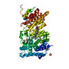
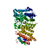
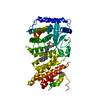
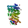
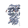
| Entry | Database: PDB / ID: 5ddf | |||||||||||||||||||||||||||
|---|---|---|---|---|---|---|---|---|---|---|---|---|---|---|---|---|---|---|---|---|---|---|---|---|---|---|---|---|
| Title | Menin in complex with MI-273 | |||||||||||||||||||||||||||
 Components Components | Menin | |||||||||||||||||||||||||||
 Keywords Keywords | PROTEIN BINDING/INHIBITOR / PROTEIN BINDING / PROTEIN BINDING-INHIBITOR complex | |||||||||||||||||||||||||||
| Function / homology |  Function and homology information Function and homology informationnegative regulation of cyclin-dependent protein serine/threonine kinase activity / Y-form DNA binding / MLL1/2 complex / osteoblast development / T-helper 2 cell differentiation / negative regulation of JNK cascade / Formation of WDR5-containing histone-modifying complexes / positive regulation of transforming growth factor beta receptor signaling pathway / negative regulation of protein phosphorylation / histone methyltransferase complex ...negative regulation of cyclin-dependent protein serine/threonine kinase activity / Y-form DNA binding / MLL1/2 complex / osteoblast development / T-helper 2 cell differentiation / negative regulation of JNK cascade / Formation of WDR5-containing histone-modifying complexes / positive regulation of transforming growth factor beta receptor signaling pathway / negative regulation of protein phosphorylation / histone methyltransferase complex / cleavage furrow / R-SMAD binding / negative regulation of cell cycle / MLL1 complex / : / negative regulation of osteoblast differentiation / response to UV / RHO GTPases activate IQGAPs / four-way junction DNA binding / transcription repressor complex / transcription initiation-coupled chromatin remodeling / response to gamma radiation / Post-translational protein phosphorylation / Deactivation of the beta-catenin transactivating complex / phosphoprotein binding / SMAD2/SMAD3:SMAD4 heterotrimer regulates transcription / Formation of the beta-catenin:TCF transactivating complex / Regulation of Insulin-like Growth Factor (IGF) transport and uptake by Insulin-like Growth Factor Binding Proteins (IGFBPs) / nuclear matrix / MAPK cascade / double-stranded DNA binding / protein-macromolecule adaptor activity / chromosome, telomeric region / transcription cis-regulatory region binding / endoplasmic reticulum lumen / negative regulation of cell population proliferation / DNA repair / negative regulation of DNA-templated transcription / chromatin binding / DNA damage response / regulation of transcription by RNA polymerase II / chromatin / negative regulation of transcription by RNA polymerase II / positive regulation of transcription by RNA polymerase II / protein-containing complex / nucleoplasm / nucleus / cytoplasm / cytosol Similarity search - Function | |||||||||||||||||||||||||||
| Biological species |  Homo sapiens (human) Homo sapiens (human) | |||||||||||||||||||||||||||
| Method |  X-RAY DIFFRACTION / SYNCHROTRON / MOLECULAR REPLACEMENT / Resolution: 1.66 Å X-RAY DIFFRACTION / SYNCHROTRON / MOLECULAR REPLACEMENT / Resolution: 1.66 Å | |||||||||||||||||||||||||||
 Authors Authors | Pollock, J. / Dmitry, B. / Cierpicki, T. / Grembecka, J. | |||||||||||||||||||||||||||
| Funding support |  United States, 8items United States, 8items
| |||||||||||||||||||||||||||
 Citation Citation | Journal: J.Med.Chem. / Year: 2015 Title: Rational Design of Orthogonal Multipolar Interactions with Fluorine in Protein-Ligand Complexes. Authors: Pollock, J. / Borkin, D. / Lund, G. / Purohit, T. / Dyguda-Kazimierowicz, E. / Grembecka, J. / Cierpicki, T. | |||||||||||||||||||||||||||
| History |
|
- Structure visualization
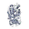
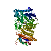
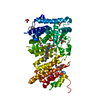
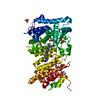
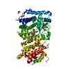
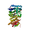
| Structure viewer | Molecule: MolmilJmol/JSmol |
|---|
- Downloads & links
-Download
| PDBx/mmCIF format | 5ddf.cif.gz | 123.3 KB | Display | PDBx/mmCIF format |
|---|---|---|---|---|
| PDB format | pdb5ddf.ent.gz | 89.7 KB | Display | PDB format |
| PDBx/mmJSON format | 5ddf.json.gz | Tree view | PDBx/mmJSON format | |
| Others |  Other downloads Other downloads |
-Validation report
| Arichive directory | https://data.pdbj.org/pub/pdb/validation_reports/dd/5ddfftp://data.pdbj.org/pub/pdb/validation_reports/dd/5ddf | HTTPS FTP |
|---|
-Related structure data
| Related structure data |  5dd9C  5ddaC 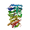 5ddbC  5ddcC  5dddC 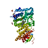 5ddeC 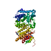 4gpqS C: citing same article ( S: Starting model for refinement |
|---|---|
| Similar structure data |


-Links
 PDBj
PDBj


- Assembly
| Deposited unit | 
| ||||||||
|---|---|---|---|---|---|---|---|---|---|
| 1 |
| ||||||||
| Unit cell |
|
-Components
-Protein , 1 types, 1 molecules A
| #1: Protein | Mass: 54570.223 Da / Num. of mol.: 1 / Fragment: UNP residues 1-459, 537-593 / Mutation: A541T Source method: isolated from a genetically manipulated source Source: (gene. exp.) Homo sapiens (human) / Gene: MEN1, SCG2 / Production host:  |
|---|
-Non-polymers , 6 types, 465 molecules 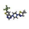


| #2: Chemical | ChemComp-5A1 /  Mass: 451.480 Da / Num. of mol.: 1 / Source method: obtained synthetically / Formula: C17H18F5N5S2 Mass: 451.480 Da / Num. of mol.: 1 / Source method: obtained synthetically / Formula: C17H18F5N5S2 | ||||||||
|---|---|---|---|---|---|---|---|---|---|
| #3: Chemical | ChemComp-DMS /  Mass: 78.133 Da / Num. of mol.: 4 / Source method: obtained synthetically / Formula: C2H6OS / Comment: DMSO, precipitant*YM Mass: 78.133 Da / Num. of mol.: 4 / Source method: obtained synthetically / Formula: C2H6OS / Comment: DMSO, precipitant*YM#4: Chemical | ChemComp-PG4 / |  Mass: 194.226 Da / Num. of mol.: 1 / Source method: obtained synthetically / Formula: C8H18O5 / Comment: precipitant*YM Mass: 194.226 Da / Num. of mol.: 1 / Source method: obtained synthetically / Formula: C8H18O5 / Comment: precipitant*YM#5: Chemical | ChemComp-PEG / |  Mass: 106.120 Da / Num. of mol.: 1 / Source method: obtained synthetically / Formula: C4H10O3 Mass: 106.120 Da / Num. of mol.: 1 / Source method: obtained synthetically / Formula: C4H10O3#6: Chemical |  Mass: 96.063 Da / Num. of mol.: 3 / Source method: obtained synthetically / Formula: SO4 Mass: 96.063 Da / Num. of mol.: 3 / Source method: obtained synthetically / Formula: SO4#7: Water | ChemComp-HOH / | Mass: 18.015 Da / Num. of mol.: 455 / Source method: isolated from a natural source / Formula: H2O |
-Experimental details
-Experiment
| Experiment | Method: X-RAY DIFFRACTION / Number of used crystals: 1 |
|---|
- Sample preparation
| Crystal | Density Matthews: 2.23 Å3/Da / Density % sol: 44.85 % |
|---|---|
| Crystal grow | Temperature: 283 K / Method: vapor diffusion, sitting drop / pH: 8 Details: 0.2 M ammonium acetate, 0.1 M HEPES and 25% w/v PEG 3,350. This solution was mixed 1:1 with 2.5mg/mL protein in 50mM Tris-HCl, 50mM NaCl, and 1mM TCEP. Prior to data collection, crystals ...Details: 0.2 M ammonium acetate, 0.1 M HEPES and 25% w/v PEG 3,350. This solution was mixed 1:1 with 2.5mg/mL protein in 50mM Tris-HCl, 50mM NaCl, and 1mM TCEP. Prior to data collection, crystals were transferred into a cryo-solution containing 20% PEG550 MME and flash-frozen in liquid nitrogen |
-Data collection
| Diffraction | Mean temperature: 100 K | ||||||||||||||||||||||||||||||||||||||||||||||||||||||||||||||||||||||||||||||||||||||||||||||||||||||||||||||||||||||||||||||
|---|---|---|---|---|---|---|---|---|---|---|---|---|---|---|---|---|---|---|---|---|---|---|---|---|---|---|---|---|---|---|---|---|---|---|---|---|---|---|---|---|---|---|---|---|---|---|---|---|---|---|---|---|---|---|---|---|---|---|---|---|---|---|---|---|---|---|---|---|---|---|---|---|---|---|---|---|---|---|---|---|---|---|---|---|---|---|---|---|---|---|---|---|---|---|---|---|---|---|---|---|---|---|---|---|---|---|---|---|---|---|---|---|---|---|---|---|---|---|---|---|---|---|---|---|---|---|---|
| Diffraction source | Source: SYNCHROTRON / Site: APS / Beamline: 21-ID-D / Wavelength: 0.97928 Å | ||||||||||||||||||||||||||||||||||||||||||||||||||||||||||||||||||||||||||||||||||||||||||||||||||||||||||||||||||||||||||||||
| Detector | Type: MARMOSAIC 300 mm CCD / Detector: CCD / Date: Aug 11, 2012 | ||||||||||||||||||||||||||||||||||||||||||||||||||||||||||||||||||||||||||||||||||||||||||||||||||||||||||||||||||||||||||||||
| Radiation | Protocol: SINGLE WAVELENGTH / Monochromatic (M) / Laue (L): M / Scattering type: x-ray | ||||||||||||||||||||||||||||||||||||||||||||||||||||||||||||||||||||||||||||||||||||||||||||||||||||||||||||||||||||||||||||||
| Radiation wavelength | Wavelength: 0.97928 Å / Relative weight: 1 | ||||||||||||||||||||||||||||||||||||||||||||||||||||||||||||||||||||||||||||||||||||||||||||||||||||||||||||||||||||||||||||||
| Reflection | Resolution: 1.66→50 Å / Num. obs: 58205 / % possible obs: 99.5 % / Redundancy: 5.9 % / Rmerge(I) obs: 0.116 / Χ2: 1.501 / Net I/av σ(I): 18.943 / Net I/σ(I): 7.2 / Num. measured all: 342112 | ||||||||||||||||||||||||||||||||||||||||||||||||||||||||||||||||||||||||||||||||||||||||||||||||||||||||||||||||||||||||||||||
| Reflection shell | Diffraction-ID: 1 / Rejects: _
|
- Processing
| Software |
| |||||||||||||||||||||||||||||||||||||||||||||||||||||||||||||||||||||||||||
|---|---|---|---|---|---|---|---|---|---|---|---|---|---|---|---|---|---|---|---|---|---|---|---|---|---|---|---|---|---|---|---|---|---|---|---|---|---|---|---|---|---|---|---|---|---|---|---|---|---|---|---|---|---|---|---|---|---|---|---|---|---|---|---|---|---|---|---|---|---|---|---|---|---|---|---|---|
| Refinement | Method to determine structure: MOLECULAR REPLACEMENT Starting model: 4GPQ Resolution: 1.66→49.34 Å / Cor.coef. Fo:Fc: 0.966 / Cor.coef. Fo:Fc free: 0.954 / Cross valid method: THROUGHOUT / σ(F): 0 / ESU R: 0.089 / ESU R Free: 0.088 / Stereochemistry target values: MAXIMUM LIKELIHOOD Details: HYDROGENS HAVE BEEN ADDED IN THE RIDING POSITIONS U VALUES : REFINED INDIVIDUALLY
| |||||||||||||||||||||||||||||||||||||||||||||||||||||||||||||||||||||||||||
| Solvent computation | Ion probe radii: 0.8 Å / Shrinkage radii: 0.8 Å / VDW probe radii: 1.2 Å / Solvent model: MASK | |||||||||||||||||||||||||||||||||||||||||||||||||||||||||||||||||||||||||||
| Displacement parameters | Biso max: 181.6 Å2 / Biso mean: 19.443 Å2 / Biso min: 5.34 Å2
| |||||||||||||||||||||||||||||||||||||||||||||||||||||||||||||||||||||||||||
| Refinement step | Cycle: final / Resolution: 1.66→49.34 Å
| |||||||||||||||||||||||||||||||||||||||||||||||||||||||||||||||||||||||||||
| Refine LS restraints |
| |||||||||||||||||||||||||||||||||||||||||||||||||||||||||||||||||||||||||||
| LS refinement shell | Resolution: 1.66→1.703 Å / Total num. of bins used: 20
|
