 Movie
Movie Controller

[English] 日本語
 Yorodumi
- PDB-6tqb: X-ray structure of Roquin ROQ domain in complex with a UCP3 CDE1 ... -
+ Open data

- Basic information
| Entry | Database: PDB / ID: 6tqb | ||||||
|---|---|---|---|---|---|---|---|
| Title | X-ray structure of Roquin ROQ domain in complex with a UCP3 CDE1 SL RNA motif | ||||||
 Components Components |
| ||||||
 Keywords Keywords | RNA BINDING PROTEIN / roquin / ROQ domain / RNA binding / UCP1 | ||||||
| Function / homology |  Function and homology information Function and homology informationnegative regulation of germinal center formation / negative regulation of T-helper cell differentiation / regulation of nuclear-transcribed mRNA catabolic process, deadenylation-dependent decay / regulation of T cell receptor signaling pathway / regulation of miRNA metabolic process / CCR4-NOT complex binding / negative regulation of T-helper 17 cell differentiation / positive regulation of mRNA catabolic process / 3'-UTR-mediated mRNA destabilization / T follicular helper cell differentiation ...negative regulation of germinal center formation / negative regulation of T-helper cell differentiation / regulation of nuclear-transcribed mRNA catabolic process, deadenylation-dependent decay / regulation of T cell receptor signaling pathway / regulation of miRNA metabolic process / CCR4-NOT complex binding / negative regulation of T-helper 17 cell differentiation / positive regulation of mRNA catabolic process / 3'-UTR-mediated mRNA destabilization / T follicular helper cell differentiation / regulation of germinal center formation / T cell proliferation / nuclear-transcribed mRNA catabolic process, nonsense-mediated decay / P-body assembly / miRNA binding / post-transcriptional regulation of gene expression / nuclear-transcribed mRNA catabolic process, deadenylation-dependent decay / negative regulation of B cell proliferation / negative regulation of activated T cell proliferation / B cell homeostasis / T cell homeostasis / lymph node development / cellular response to interleukin-1 / spleen development / nuclear-transcribed mRNA catabolic process / regulation of mRNA stability / mRNA 3'-UTR binding / P-body / positive regulation of non-canonical NF-kappaB signal transduction / RING-type E3 ubiquitin transferase / RNA stem-loop binding / cytoplasmic stress granule / protein polyubiquitination / ubiquitin-protein transferase activity / ubiquitin protein ligase activity / T cell receptor signaling pathway / double-stranded RNA binding / regulation of gene expression / ubiquitin-dependent protein catabolic process / mRNA binding / zinc ion binding / cytoplasm Similarity search - Function | ||||||
| Biological species |  | ||||||
| Method |  X-RAY DIFFRACTION / SYNCHROTRON / MOLECULAR REPLACEMENT / Resolution: 1.6 Å X-RAY DIFFRACTION / SYNCHROTRON / MOLECULAR REPLACEMENT / Resolution: 1.6 Å | ||||||
 Authors Authors | Binas, O. / Tants, J.-N. / Peter, S.A. / Janowski, R. / Davydova, E. / Braun, J. / Niessing, D. / Schwalbe, H. / Weigand, J.E. / Schlundt, A. | ||||||
 Citation Citation | Journal: Nucleic Acids Res. / Year: 2020 Title: Structural basis for the recognition of transiently structured AU-rich elements by Roquin. Authors: Binas, O. / Tants, J.N. / Peter, S.A. / Janowski, R. / Davydova, E. / Braun, J. / Niessing, D. / Schwalbe, H. / Weigand, J.E. / Schlundt, A. | ||||||
| History |
|
- Structure visualization
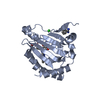
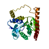
| Structure viewer | Molecule: MolmilJmol/JSmol |
|---|
- Downloads & links
-Download
| PDBx/mmCIF format | 6tqb.cif.gz | 73.3 KB | Display | PDBx/mmCIF format |
|---|---|---|---|---|
| PDB format | pdb6tqb.ent.gz | 50.5 KB | Display | PDB format |
| PDBx/mmJSON format | 6tqb.json.gz | Tree view | PDBx/mmJSON format | |
| Others |  Other downloads Other downloads |
-Validation report
| Arichive directory | https://data.pdbj.org/pub/pdb/validation_reports/tq/6tqbftp://data.pdbj.org/pub/pdb/validation_reports/tq/6tqb | HTTPS FTP |
|---|
-Related structure data
| Related structure data |  6tqaC  6xwjC 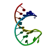 6xwwC  6xxaC  6xxbC  4qi2S S: Starting model for refinement C: citing same article ( |
|---|---|
| Similar structure data |


-Links
 PDBj
PDBj


- Assembly
| Deposited unit | 
| ||||||||
|---|---|---|---|---|---|---|---|---|---|
| 1 |
| ||||||||
| Unit cell |
| ||||||||
| Components on special symmetry positions |
|
-Components
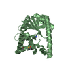
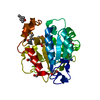
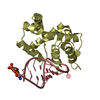
-Protein / RNA chain , 2 types, 2 molecules AB
| #1: Protein | Mass: 20528.561 Da / Num. of mol.: 1 Source method: isolated from a genetically manipulated source Source: (gene. exp.)  References: UniProt: Q4VGL6, RING-type E3 ubiquitin transferase |
|---|---|
| #2: RNA chain | Mass: 6009.584 Da / Num. of mol.: 1 / Source method: obtained synthetically / Source: (synth.) |
-Non-polymers , 5 types, 252 molecules 


| #3: Chemical | ChemComp-EDO /  Mass: 62.068 Da / Num. of mol.: 6 / Source method: obtained synthetically / Formula: C2H6O2 Mass: 62.068 Da / Num. of mol.: 6 / Source method: obtained synthetically / Formula: C2H6O2#4: Chemical |  Mass: 22.990 Da / Num. of mol.: 2 / Source method: obtained synthetically / Formula: Na Mass: 22.990 Da / Num. of mol.: 2 / Source method: obtained synthetically / Formula: Na#5: Chemical |  Mass: 35.453 Da / Num. of mol.: 2 / Source method: obtained synthetically / Formula: Cl Mass: 35.453 Da / Num. of mol.: 2 / Source method: obtained synthetically / Formula: Cl#6: Chemical | ChemComp-MG / |  Mass: 24.305 Da / Num. of mol.: 1 / Source method: obtained synthetically / Formula: Mg Mass: 24.305 Da / Num. of mol.: 1 / Source method: obtained synthetically / Formula: Mg#7: Water | ChemComp-HOH / | Mass: 18.015 Da / Num. of mol.: 241 / Source method: isolated from a natural source / Formula: H2O |
|---|
-Details
| Has ligand of interest | N |
|---|
-Experimental details
-Experiment
| Experiment | Method: X-RAY DIFFRACTION / Number of used crystals: 1 |
|---|
- Sample preparation
| Crystal | Density Matthews: 2.6 Å3/Da / Density % sol: 52.72 % |
|---|---|
| Crystal grow | Temperature: 292 K / Method: vapor diffusion, hanging drop Details: 0.01 M Bis Tris Propane pH 6.5, 0.29 M Sodium Tartrate, 19 % PEG 3350 |
-Data collection
| Diffraction | Mean temperature: 100 K / Serial crystal experiment: N |
|---|---|
| Diffraction source | Source: SYNCHROTRON / Site: SLS  / Beamline: X06DA / Wavelength: 1 Å / Beamline: X06DA / Wavelength: 1 Å |
| Detector | Type: DECTRIS PILATUS 2M-F / Detector: PIXEL / Date: Jul 14, 2019 |
| Radiation | Protocol: SINGLE WAVELENGTH / Monochromatic (M) / Laue (L): M / Scattering type: x-ray |
| Radiation wavelength | Wavelength: 1 Å / Relative weight: 1 |
| Reflection | Resolution: 1.6→50 Å / Num. obs: 36355 / % possible obs: 96.9 % / Observed criterion σ(F): 0 / Observed criterion σ(I): 0 / Redundancy: 8.4 % / Biso Wilson estimate: 31 Å2 / CC1/2: 1 / Rmerge(I) obs: 0.047 / Net I/σ(I): 23.16 |
| Reflection shell | Resolution: 1.6→1.64 Å / Redundancy: 6.3 % / Rmerge(I) obs: 0.736 / Mean I/σ(I) obs: 2.46 / Num. unique obs: 2053 / CC1/2: 0.797 / % possible all: 75.5 |
- Processing
| Software |
| ||||||||||||||||||||||||||||||||||||||||||||||||||||||||||||
|---|---|---|---|---|---|---|---|---|---|---|---|---|---|---|---|---|---|---|---|---|---|---|---|---|---|---|---|---|---|---|---|---|---|---|---|---|---|---|---|---|---|---|---|---|---|---|---|---|---|---|---|---|---|---|---|---|---|---|---|---|---|
| Refinement | Method to determine structure: MOLECULAR REPLACEMENT Starting model: 4QI2 Resolution: 1.6→47.08 Å / Cor.coef. Fo:Fc: 0.975 / Cor.coef. Fo:Fc free: 0.96 / SU B: 1.482 / SU ML: 0.051 / Cross valid method: THROUGHOUT / σ(F): 0 / ESU R: 0.075 / ESU R Free: 0.082 / Stereochemistry target values: MAXIMUM LIKELIHOOD Details: HYDROGENS HAVE BEEN ADDED IN THE RIDING POSITIONS U VALUES : REFINED INDIVIDUALLY
| ||||||||||||||||||||||||||||||||||||||||||||||||||||||||||||
| Solvent computation | Ion probe radii: 0.8 Å / Shrinkage radii: 0.8 Å / VDW probe radii: 1.2 Å / Solvent model: MASK | ||||||||||||||||||||||||||||||||||||||||||||||||||||||||||||
| Displacement parameters | Biso max: 143.37 Å2 / Biso mean: 28.248 Å2 / Biso min: 16.39 Å2
| ||||||||||||||||||||||||||||||||||||||||||||||||||||||||||||
| Refinement step | Cycle: final / Resolution: 1.6→47.08 Å
| ||||||||||||||||||||||||||||||||||||||||||||||||||||||||||||
| Refine LS restraints |
| ||||||||||||||||||||||||||||||||||||||||||||||||||||||||||||
| LS refinement shell | Resolution: 1.6→1.642 Å / Rfactor Rfree error: 0 / Total num. of bins used: 20
|
