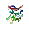
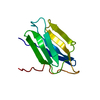
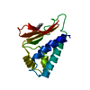
Zinc finger C2H2 type domain profile. / Zinc finger C2H2-type / Serine/threonine-protein kinase, active site / Serine/Threonine protein kinases active-site signature. / Protein kinase domain / Serine/Threonine protein kinases, catalytic domain / Protein kinase, ATP binding site / Protein kinases ATP-binding region signature. / Protein kinase domain profile. / Protein kinase domain / Protein kinase-like domain superfamily Similarity search - Domain/homology
Protocol: SINGLE WAVELENGTH / Monochromatic (M) / Laue (L): M / Scattering type: x-ray
Radiation wavelength
Wavelength: 1.28297 Å / Relative weight: 1
Reflection
Resolution: 1.8→45.02 Å / Num. obs: 8158 / % possible obs: 97.1 % / Redundancy: 4.6 % / Biso Wilson estimate: 29.18 Å2 / CC1/2: 0.99 / Rmerge(I) obs: 0.102 / Rpim(I) all: 0.05 / Net I/σ(I): 8.7
Reflection shell
Diffraction-ID: 1
Resolution (Å)
Redundancy (%)
Rmerge(I) obs
Num. unique obs
CC1/2
Rpim(I) all
% possible all
1.8-1.83
2.8
0.52
334
0.78
0.33
77.9
4.88-45.04
4.2
0.072
459
0.99
0.035
97.9
-
Phasing
Phasing
Method: SAD
-
Processing
Software
Name
Version
Classification
Aimless
datascaling
BUSTER
2.10.3 (3-OCT-2019)
refinement
PDB_EXTRACT
3.25
dataextraction
DIALS
datareduction
CRANK2
phasing
Refinement
Method to determine structure: SAD / Resolution: 1.8→35 Å / Cor.coef. Fo:Fc: 0.949 / Cor.coef. Fo:Fc free: 0.927 / SU R Cruickshank DPI: 0.152 / Cross valid method: THROUGHOUT / σ(F): 0 / SU R Blow DPI: 0.176 / SU Rfree Blow DPI: 0.143 / SU Rfree Cruickshank DPI: 0.133
In the structure databanks used in Yorodumi, some data are registered as the other names, "COVID-19 virus" and "2019-nCoV". Here are the details of the virus and the list of structure data.
Jan 31, 2019. EMDB accession codes are about to change! (news from PDBe EMDB page)
EMDB accession codes are about to change! (news from PDBe EMDB page)
The allocation of 4 digits for EMDB accession codes will soon come to an end. Whilst these codes will remain in use, new EMDB accession codes will include an additional digit and will expand incrementally as the available range of codes is exhausted. The current 4-digit format prefixed with “EMD-” (i.e. EMD-XXXX) will advance to a 5-digit format (i.e. EMD-XXXXX), and so on. It is currently estimated that the 4-digit codes will be depleted around Spring 2019, at which point the 5-digit format will come into force.
The EM Navigator/Yorodumi systems omit the EMD- prefix.
Related info.:Q: What is EMD? / ID/Accession-code notation in Yorodumi/EM Navigator
Yorodumi is a browser for structure data from EMDB, PDB, SASBDB, etc.
This page is also the successor to EM Navigator detail page, and also detail information page/front-end page for Omokage search.
The word "yorodu" (or yorozu) is an old Japanese word meaning "ten thousand". "mi" (miru) is to see.
Related info.:EMDB / PDB / SASBDB / Comparison of 3 databanks / Yorodumi Search / Aug 31, 2016. New EM Navigator & Yorodumi / Yorodumi Papers / Jmol/JSmol / Function and homology information / Changes in new EM Navigator and Yorodumi
 Movie
Movie Controller

 Yorodumi
 Open data
 Basic information
 Components
 Keywords
 Function and homology information
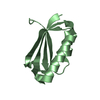
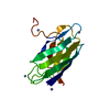
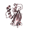
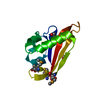
 Bodo saltans (eukaryote)
 X-RAY DIFFRACTION /
  Authors
 United Kingdom, 1items
  Citation
 Structure visualization
 Downloads & links
 Other downloads
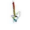


 PDBj
PDBj


 Assembly


 Mass: 65.409 Da / Num. of mol.: 3 / Source method: obtained synthetically / Formula: Zn / Feature type: SUBJECT OF INVESTIGATION
 Mass: 96.063 Da / Num. of mol.: 2 / Source method: obtained synthetically / Formula: SO4
 Mass: 35.453 Da / Num. of mol.: 1 / Source method: obtained synthetically / Formula: Cl
 Mass: 18.015 Da / Num. of mol.: 94 / Source method: isolated from a natural source / Formula: H2O
 Sample preparation
 Processing