 Movie
Movie Controller

[English] 日本語
 Yorodumi
- PDB-6tlx: Crystal structure of the unconventional kinetochore protein Perki... -
+ Open data

- Basic information
| Entry | Database: PDB / ID: 6tlx | ||||||
|---|---|---|---|---|---|---|---|
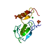
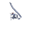
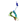
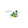
| Title | Crystal structure of the unconventional kinetochore protein Perkinsela sp. KKT2a central domain | ||||||
 Components Components | Protein kinase | ||||||
 Keywords Keywords | CELL CYCLE / kinetochore / zinc finger / kinetoplastid / KKT2 | ||||||
| Function / homology |  Function and homology information Function and homology information | ||||||
| Biological species |  Perkinsela sp. CCAP 1560/4 (eukaryote) Perkinsela sp. CCAP 1560/4 (eukaryote) | ||||||
| Method |  X-RAY DIFFRACTION / SYNCHROTRON / SAD / Resolution: 2.87 Å X-RAY DIFFRACTION / SYNCHROTRON / SAD / Resolution: 2.87 Å | ||||||
 Authors Authors | Marciano, G. / Nerusheva, O. / Ishii, M. / Akiyoshi, B. | ||||||
| Funding support |  United Kingdom, 1items United Kingdom, 1items
| ||||||
 Citation Citation | Journal: J.Cell Biol. / Year: 2021 Title: Kinetoplastid kinetochore proteins KKT2 and KKT3 have unique centromere localization domains. Authors: Marciano, G. / Ishii, M. / Nerusheva, O.O. / Akiyoshi, B. #1: Journal: Biorxiv / Year: 2019Title: Unconventional kinetochore kinases KKT2 and KKT3 have a unique zinc finger that promotes their kinetochore localization Authors: Marciano, G. / Nerusheva, O. / Ishii, M. / Akiyoshi, B. | ||||||
| History |
|
- Structure visualization
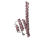
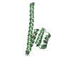
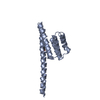
| Structure viewer | Molecule: MolmilJmol/JSmol |
|---|
- Downloads & links
-Download
| PDBx/mmCIF format | 6tlx.cif.gz | 36.7 KB | Display | PDBx/mmCIF format |
|---|---|---|---|---|
| PDB format | pdb6tlx.ent.gz | 22.7 KB | Display | PDB format |
| PDBx/mmJSON format | 6tlx.json.gz | Tree view | PDBx/mmJSON format | |
| Others |  Other downloads Other downloads |
-Validation report
| Arichive directory | https://data.pdbj.org/pub/pdb/validation_reports/tl/6tlxftp://data.pdbj.org/pub/pdb/validation_reports/tl/6tlx | HTTPS FTP |
|---|
-Related structure data

-Links
 PDBj
PDBj
- Assembly
| Deposited unit | 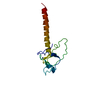
| ||||||||
|---|---|---|---|---|---|---|---|---|---|
| 1 |
| ||||||||
| Unit cell |
|
-Components
| #1: Protein | Mass: 14570.530 Da / Num. of mol.: 1 Source method: isolated from a genetically manipulated source Source: (gene. exp.) Perkinsela sp. CCAP 1560/4 (eukaryote) / Gene: XU18_4017 / Plasmid: RSFDuet-1 / Production host:  | ||
|---|---|---|---|
| #2: Chemical |   Mass: 65.409 Da / Num. of mol.: 2 / Source method: obtained synthetically / Formula: Zn / Feature type: SUBJECT OF INVESTIGATION Mass: 65.409 Da / Num. of mol.: 2 / Source method: obtained synthetically / Formula: Zn / Feature type: SUBJECT OF INVESTIGATIONHas ligand of interest | Y | |
-Experimental details
-Experiment
| Experiment | Method: X-RAY DIFFRACTION / Number of used crystals: 1 |
|---|
- Sample preparation
| Crystal | Density Matthews: 5.91 Å3/Da / Density % sol: 79.18 % |
|---|---|
| Crystal grow | Temperature: 277 K / Method: vapor diffusion, sitting drop / pH: 7.5 / Details: 19% MPD, 50 mM Hepes, pH 7.5, 10 mM MgCl2 |
-Data collection
| Diffraction | Mean temperature: 100 K / Serial crystal experiment: N | ||||||||||||||||||||||||
|---|---|---|---|---|---|---|---|---|---|---|---|---|---|---|---|---|---|---|---|---|---|---|---|---|---|
| Diffraction source | Source: SYNCHROTRON / Site: Diamond / Beamline: I24 / Wavelength: 0.9686 Å | ||||||||||||||||||||||||
| Detector | Type: DECTRIS PILATUS3 6M / Detector: PIXEL / Date: Feb 12, 2018 | ||||||||||||||||||||||||
| Radiation | Protocol: SINGLE WAVELENGTH / Monochromatic (M) / Laue (L): M / Scattering type: x-ray | ||||||||||||||||||||||||
| Radiation wavelength | Wavelength: 0.9686 Å / Relative weight: 1 | ||||||||||||||||||||||||
| Reflection | Resolution: 2.87→56.92 Å / Num. obs: 7986 / % possible obs: 99.7 % / Redundancy: 19.2 % / Biso Wilson estimate: 105.112 Å2 / CC1/2: 1 / Rmerge(I) obs: 0.102 / Rpim(I) all: 0.024 / Rrim(I) all: 0.105 / Net I/σ(I): 16.4 | ||||||||||||||||||||||||
| Reflection shell | Diffraction-ID: 1
|
-Phasing
| Phasing | Method: SAD |
|---|
- Processing
| Software |
| ||||||||||||||||||||||||||||||||||||||||||||||||||||||||||||||||||||||||||||||||||||||||||||||||||||||||||||
|---|---|---|---|---|---|---|---|---|---|---|---|---|---|---|---|---|---|---|---|---|---|---|---|---|---|---|---|---|---|---|---|---|---|---|---|---|---|---|---|---|---|---|---|---|---|---|---|---|---|---|---|---|---|---|---|---|---|---|---|---|---|---|---|---|---|---|---|---|---|---|---|---|---|---|---|---|---|---|---|---|---|---|---|---|---|---|---|---|---|---|---|---|---|---|---|---|---|---|---|---|---|---|---|---|---|---|---|---|---|
| Refinement | Method to determine structure: SAD / Resolution: 2.87→49 Å / Cor.coef. Fo:Fc: 0.942 / Cor.coef. Fo:Fc free: 0.916 / SU R Cruickshank DPI: 0.315 / Cross valid method: THROUGHOUT / σ(F): 0 / SU R Blow DPI: 0.306 / SU Rfree Blow DPI: 0.253 / SU Rfree Cruickshank DPI: 0.259
| ||||||||||||||||||||||||||||||||||||||||||||||||||||||||||||||||||||||||||||||||||||||||||||||||||||||||||||
| Displacement parameters | Biso max: 163.08 Å2 / Biso mean: 113.37 Å2 / Biso min: 87.09 Å2
| ||||||||||||||||||||||||||||||||||||||||||||||||||||||||||||||||||||||||||||||||||||||||||||||||||||||||||||
| Refine analyze | Luzzati coordinate error obs: 0.56 Å | ||||||||||||||||||||||||||||||||||||||||||||||||||||||||||||||||||||||||||||||||||||||||||||||||||||||||||||
| Refinement step | Cycle: final / Resolution: 2.87→49 Å
| ||||||||||||||||||||||||||||||||||||||||||||||||||||||||||||||||||||||||||||||||||||||||||||||||||||||||||||
| Refine LS restraints |
| ||||||||||||||||||||||||||||||||||||||||||||||||||||||||||||||||||||||||||||||||||||||||||||||||||||||||||||
| LS refinement shell | Resolution: 2.87→3 Å / Rfactor Rfree error: 0 / Total num. of bins used: 18
|
