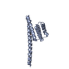
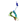
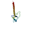
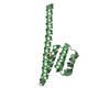
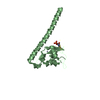
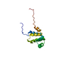
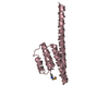
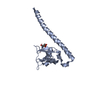
Neurabin-2 / Neurabin-II / Neural tissue-specific F-actin binding protein II / Protein phosphatase 1 regulatory ...Neurabin-II / Neural tissue-specific F-actin binding protein II / Protein phosphatase 1 regulatory subunit 9B / Spinophilin / p130 / PP1bp134
Mass: 12135.745 Da / Num. of mol.: 1 / Fragment: PDZ domain Source method: isolated from a genetically manipulated source Source: (gene. exp.) Rattus norvegicus (Norway rat) / Gene: Ppp1r9b / Plasmid: derivitive of pET28a / Production host: Escherichia coli (E. coli) / Strain (production host): BL21 (DE3) RIL codon plus / References: UniProt: O35274
-
Experimental details
-
Experiment
Experiment
Method: SOLUTION NMR
NMR experiment
Conditions-ID
Experiment-ID
Solution-ID
Type
1
1
1
3D 15N resolved [1H,1H] NOESY
1
2
2
3D 13C resolved [1H,1H] NOESY
1
3
3
2D 1H-1H NOESY
NMR details
Text: The following spectra were used to achieve the sequence-specific backbone and side chain assignments of all aliphatic residues: 2D [1H,15N]-HSQC, 2D [1H,13C]-HSQC, 3D HNCACB, 3D CBCA(CO)NH, 3D ...Text: The following spectra were used to achieve the sequence-specific backbone and side chain assignments of all aliphatic residues: 2D [1H,15N]-HSQC, 2D [1H,13C]-HSQC, 3D HNCACB, 3D CBCA(CO)NH, 3D CC(CO)NH, 3D HNCO, 3D HNCA, 3D HBHA(CO)NH, 3D 15N-resolved [1H,1H]-TOCSY, 3D HC(C)H-TOCSY6. The 2D [1H,1H]-NOESY, 2D [1H,1H]-TOCSY and 2D [1H,1H]-COSY spectra of the Spinophilin493-602 sample in D2O solution after complete H/D exchange of the labile protons were used for the assignment of the aromatic side chains. The NMR spectra were processed with Topspin1.3, and analyzed with the CARA software package. Spectra used for the structure calculation were: 3D 15N-resolved [1H,1H]-NOESY, 3D 13C-resolved [1H,1H]-NOESY (mixing time of 85 ms) and 2D [1H,1H]-NOESY (mixing time 85 ms, D2O solution).
Ionic strength: 20 mM Na phosphate; 50 mM NaCl / pH: 6.5 / Pressure: ambient / Temperature: 298 K
-
NMR measurement
Radiation
Protocol: SINGLE WAVELENGTH / Monochromatic (M) / Laue (L): M
Radiation wavelength
Relative weight: 1
NMR spectrometer
Type: Bruker AVANCE / Manufacturer: Bruker / Model: AVANCE / Field strength: 500 MHz
-
Processing
NMR software
Name
Version
Developer
Classification
TopSpin
1.3
Bruker
processing
CYANA
2
peterguntert
structuresolution
CNS
1.1
Brunger
refinement
atnos/candid
2
TorstenHerrmann
structuresolution
CARA
1.5
RochusKeller
dataanalysis
Refinement
Method: simulated annealing torsion angle dynamics / Software ordinal: 1 Details: The amino acid sequence, the chemical shift assignment, and the NOESY spectra were input for the automated NOESY peak picking and NOE assignment method of ATNOS/CANDID/CYANA. The results of ...Details: The amino acid sequence, the chemical shift assignment, and the NOESY spectra were input for the automated NOESY peak picking and NOE assignment method of ATNOS/CANDID/CYANA. The results of ATNOS/CANDID/CYANA were refined by manual peak adjustment and additional calculations in CYANA. The 20 conformers from the ATNOS/CANDID cycle 7 with the lowest residual CYANA target function values were energy-minimized in a water shell with the program CNS.
NMR representative
Selection criteria: closest to the average
NMR ensemble
Conformer selection criteria: structures with the lowest energy Conformers calculated total number: 50 / Conformers submitted total number: 20
+
About Yorodumi
-
News
-
Feb 9, 2022. New format data for meta-information of EMDB entries
New format data for meta-information of EMDB entries
Version 3 of the EMDB header file is now the official format.
The previous official version 1.9 will be removed from the archive.
In the structure databanks used in Yorodumi, some data are registered as the other names, "COVID-19 virus" and "2019-nCoV". Here are the details of the virus and the list of structure data.
Jan 31, 2019. EMDB accession codes are about to change! (news from PDBe EMDB page)
EMDB accession codes are about to change! (news from PDBe EMDB page)
The allocation of 4 digits for EMDB accession codes will soon come to an end. Whilst these codes will remain in use, new EMDB accession codes will include an additional digit and will expand incrementally as the available range of codes is exhausted. The current 4-digit format prefixed with “EMD-” (i.e. EMD-XXXX) will advance to a 5-digit format (i.e. EMD-XXXXX), and so on. It is currently estimated that the 4-digit codes will be depleted around Spring 2019, at which point the 5-digit format will come into force.
The EM Navigator/Yorodumi systems omit the EMD- prefix.
Related info.:Q: What is EMD? / ID/Accession-code notation in Yorodumi/EM Navigator
Yorodumi is a browser for structure data from EMDB, PDB, SASBDB, etc.
This page is also the successor to EM Navigator detail page, and also detail information page/front-end page for Omokage search.
The word "yorodu" (or yorozu) is an old Japanese word meaning "ten thousand". "mi" (miru) is to see.
Related info.:EMDB / PDB / SASBDB / Comparison of 3 databanks / Yorodumi Search / Aug 31, 2016. New EM Navigator & Yorodumi / Yorodumi Papers / Jmol/JSmol / Function and homology information / Changes in new EM Navigator and Yorodumi
 Movie
Movie Controller


 Open data
 Basic information
 Components
 Keywords
 Function and homology information
 Authors
 Citation
 Structure visualization
 Downloads & links
 Other downloads
 PDBj
PDBj Assembly

 Sample preparation
 Processing
 CYANA
CYANA