 Movie
Movie Controller

[English] 日本語
 Yorodumi
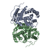
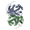
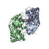
- PDB-6tja: Crystal structure of the SVS_A2 protein (W79F,G83L mutant) from a... -
+ Open data

- Basic information
| Entry | Database: PDB / ID: 6tja | ||||||
|---|---|---|---|---|---|---|---|
| Title | Crystal structure of the SVS_A2 protein (W79F,G83L mutant) from ancestral sequence reconstruction at 2.27 A resolution | ||||||
 Components Components | SVS_variant_AS1 | ||||||
 Keywords Keywords | BIOSYNTHETIC PROTEIN / Terpene cyclase / engineered enzyme | ||||||
| Function / homology | DI(HYDROXYETHYL)ETHER Function and homology information Function and homology information | ||||||
| Biological species |  Streptomyces sp. CWA1 (bacteria) Streptomyces sp. CWA1 (bacteria) | ||||||
| Method |  X-RAY DIFFRACTION / SYNCHROTRON / MOLECULAR REPLACEMENT / Resolution: 2.27 Å X-RAY DIFFRACTION / SYNCHROTRON / MOLECULAR REPLACEMENT / Resolution: 2.27 Å | ||||||
 Authors Authors | Rudraraju, R. / Schnell, R. / Schneider, G. | ||||||
| Funding support |  Sweden, 1items Sweden, 1items
| ||||||
 Citation Citation | Journal: J.Am.Chem.Soc. / Year: 2021 Title: Engineering of Ancestors as a Tool to Elucidate Structure, Mechanism, and Specificity of Extant Terpene Cyclase. Authors: Schriever, K. / Saenz-Mendez, P. / Rudraraju, R.S. / Hendrikse, N.M. / Hudson, E.P. / Biundo, A. / Schnell, R. / Syren, P.O. | ||||||
| History |
|
- Structure visualization
| Structure viewer | Molecule: MolmilJmol/JSmol |
|---|
- Downloads & links
-Download
| PDBx/mmCIF format | 6tja.cif.gz | 142.6 KB | Display | PDBx/mmCIF format |
|---|---|---|---|---|
| PDB format | pdb6tja.ent.gz | 111.4 KB | Display | PDB format |
| PDBx/mmJSON format | 6tja.json.gz | Tree view | PDBx/mmJSON format | |
| Others |  Other downloads Other downloads |
-Validation report
| Arichive directory | https://data.pdbj.org/pub/pdb/validation_reports/tj/6tjaftp://data.pdbj.org/pub/pdb/validation_reports/tj/6tja | HTTPS FTP |
|---|
-Related structure data
| Related structure data |  6tbdC  6thuC  6tivC 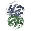 6tjzC  4okzS S: Starting model for refinement C: citing same article ( |
|---|---|
| Similar structure data |


-Links
 PDBj
PDBj- Assembly
| Deposited unit | 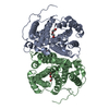
| ||||||||
|---|---|---|---|---|---|---|---|---|---|
| 1 |
| ||||||||
| Unit cell |
|
-Components
| #1: Protein | Mass: 40492.852 Da / Num. of mol.: 2 Source method: isolated from a genetically manipulated source Details: artificial protein / Source: (gene. exp.) Streptomyces sp. CWA1 (bacteria) / Production host: #2: Chemical |   Mass: 106.120 Da / Num. of mol.: 3 / Source method: obtained synthetically / Formula: C4H10O3 Mass: 106.120 Da / Num. of mol.: 3 / Source method: obtained synthetically / Formula: C4H10O3#3: Water | ChemComp-HOH / |  Mass: 18.015 Da / Num. of mol.: 218 / Source method: isolated from a natural source / Formula: H2O Mass: 18.015 Da / Num. of mol.: 218 / Source method: isolated from a natural source / Formula: H2OHas ligand of interest | N | |
|---|
-Experimental details
-Experiment
| Experiment | Method: X-RAY DIFFRACTION / Number of used crystals: 1 |
|---|
- Sample preparation
| Crystal | Density Matthews: 2.62 Å3/Da / Density % sol: 53.05 % |
|---|---|
| Crystal grow | Temperature: 293 K / Method: vapor diffusion, hanging drop / pH: 7.2 Details: 0.2M Na phosphate 0.1M Bis-Tris-Propane pH 7.2 12% PEG 3350 Cryoprotection: 0.2M Na phosphate 0.1M Bis-Tris-Propane pH 7.2 20% PEG 3350 5mM MgCl2 FPP |
-Data collection
| Diffraction | Mean temperature: 100 K / Serial crystal experiment: N |
|---|---|
| Diffraction source | Source: SYNCHROTRON / Site: MAX IV / Beamline: BioMAX / Wavelength: 0.91841 Å |
| Detector | Type: DECTRIS EIGER X 16M / Detector: PIXEL / Date: Jun 28, 2019 / Details: KB mirrors |
| Radiation | Monochromator: Si (111) / Protocol: SINGLE WAVELENGTH / Monochromatic (M) / Laue (L): M / Scattering type: x-ray |
| Radiation wavelength | Wavelength: 0.91841 Å / Relative weight: 1 |
| Reflection | Resolution: 2.27→29.43 Å / Num. obs: 40139 / % possible obs: 99.8 % / Redundancy: 5.6 % / Biso Wilson estimate: 30 Å2 / CC1/2: 0.976 / Rmerge(I) obs: 0.1 / Rpim(I) all: 0.05 / Net I/σ(I): 9.7 |
| Reflection shell | Resolution: 2.27→2.34 Å / Rmerge(I) obs: 0.528 / Mean I/σ(I) obs: 2.7 / Num. unique obs: 3637 / CC1/2: 0.853 / Rpim(I) all: 0.264 / % possible all: 99.6 |
- Processing
| Software |
| ||||||||||||||||||||||||||||||||||||||||||||||||||||||||||||
|---|---|---|---|---|---|---|---|---|---|---|---|---|---|---|---|---|---|---|---|---|---|---|---|---|---|---|---|---|---|---|---|---|---|---|---|---|---|---|---|---|---|---|---|---|---|---|---|---|---|---|---|---|---|---|---|---|---|---|---|---|---|
| Refinement | Method to determine structure: MOLECULAR REPLACEMENT Starting model: 4OKZ Resolution: 2.27→29.43 Å / Cor.coef. Fo:Fc: 0.953 / Cor.coef. Fo:Fc free: 0.932 / SU B: 5.785 / SU ML: 0.141 / Cross valid method: THROUGHOUT / σ(F): 0 / ESU R: 0.24 / ESU R Free: 0.191 Details: HYDROGENS HAVE BEEN ADDED IN THE RIDING POSITIONS U VALUES : REFINED INDIVIDUALLY
| ||||||||||||||||||||||||||||||||||||||||||||||||||||||||||||
| Solvent computation | Ion probe radii: 0.8 Å / Shrinkage radii: 0.8 Å / VDW probe radii: 1.2 Å | ||||||||||||||||||||||||||||||||||||||||||||||||||||||||||||
| Displacement parameters | Biso max: 131.69 Å2 / Biso mean: 35.637 Å2 / Biso min: 18.36 Å2
| ||||||||||||||||||||||||||||||||||||||||||||||||||||||||||||
| Refinement step | Cycle: final / Resolution: 2.27→29.43 Å
| ||||||||||||||||||||||||||||||||||||||||||||||||||||||||||||
| Refine LS restraints |
| ||||||||||||||||||||||||||||||||||||||||||||||||||||||||||||
| LS refinement shell | Resolution: 2.27→2.329 Å / Rfactor Rfree error: 0 / Total num. of bins used: 20
|
