 Movie
Movie Controller

[English] 日本語
 Yorodumi
- PDB-1ysl: Crystal structure of HMG-CoA synthase from Enterococcus faecalis ... -
+ Open data

- Basic information
| Entry | Database: PDB / ID: 1ysl | ||||||
|---|---|---|---|---|---|---|---|
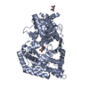
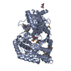
| Title | Crystal structure of HMG-CoA synthase from Enterococcus faecalis with AcetoAcetyl-CoA ligand. | ||||||
 Components Components | (HMG-CoA synthase) x 2 | ||||||
 Keywords Keywords | LYASE / Thiolase family / CoEnzymeA | ||||||
| Function / homology |  Function and homology information Function and homology informationhydroxymethylglutaryl-CoA synthase / hydroxymethylglutaryl-CoA synthase activity / acetyl-CoA metabolic process / isoprenoid biosynthetic process Similarity search - Function | ||||||
| Biological species |   Enterococcus faecalis (bacteria) Enterococcus faecalis (bacteria) | ||||||
| Method |  X-RAY DIFFRACTION / SYNCHROTRON / MOLECULAR REPLACEMENT / Resolution: 1.9 Å X-RAY DIFFRACTION / SYNCHROTRON / MOLECULAR REPLACEMENT / Resolution: 1.9 Å | ||||||
 Authors Authors | Steussy, C.N. / Vartia, A.A. / Burgner II, J.W. / Sutherlin, A. / Rodwell, V.W. / Stauffacher, C.V. | ||||||
 Citation Citation | Journal: Biochemistry / Year: 2005 Title: X-ray Crystal Structures of HMG-CoA Synthase from Enterococcus faecalis and a Complex with Its Second Substrate/Inhibitor Acetoacetyl-CoA. Authors: Steussy, C.N. / Vartia, A.A. / Burgner II, J.W. / Sutherlin, A. / Rodwell, V.W. / Stauffacher, C.V. | ||||||
| History |
| ||||||
| Remark 400 | COMPOUND THE ACETOACETYL MOLECULE, AAE IS COVALENTLY ATTACHED TO CYSTEINE B111 WITH 50% OCCUPANCY. ...COMPOUND THE ACETOACETYL MOLECULE, AAE IS COVALENTLY ATTACHED TO CYSTEINE B111 WITH 50% OCCUPANCY. THE REMAINING PORTION OF CYSTEINE B111 IS OXIDIZED TO SULFONATE (CSD). CYSTEINE A 111 IS ENTIRELY OXIDIZED (CSD). |
- Structure visualization
| Structure viewer | Molecule: MolmilJmol/JSmol |
|---|
- Downloads & links
-Download
| PDBx/mmCIF format | 1ysl.cif.gz | 176.1 KB | Display | PDBx/mmCIF format |
|---|---|---|---|---|
| PDB format | pdb1ysl.ent.gz | 137.4 KB | Display | PDB format |
| PDBx/mmJSON format | 1ysl.json.gz | Tree view | PDBx/mmJSON format | |
| Others |  Other downloads Other downloads |
-Validation report
| Arichive directory | https://data.pdbj.org/pub/pdb/validation_reports/ys/1yslftp://data.pdbj.org/pub/pdb/validation_reports/ys/1ysl | HTTPS FTP |
|---|
-Related structure data
| Related structure data |  1x9eSC S: Starting model for refinement C: citing same article ( |
|---|---|
| Similar structure data |


-Links
 PDBj
PDBj


- Assembly
| Deposited unit | 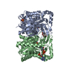
| ||||||||
|---|---|---|---|---|---|---|---|---|---|
| 1 |
| ||||||||
| Unit cell |
| ||||||||
| Components on special symmetry positions |
| ||||||||
| Details | The asymmetric unit contains a homodimer that is believed to be the biologically active unit. |
-Components
-Protein , 2 types, 2 molecules AB
| #1: Protein | Mass: 44307.852 Da / Num. of mol.: 1 Source method: isolated from a genetically manipulated source Source: (gene. exp.) Enterococcus faecalis (bacteria) / Plasmid: pET28a / Production host: References: GenBank: 9937383, UniProt: Q9FD71*PLUS, EC: 4.1.3.5 |
|---|---|
| #2: Protein | Mass: 44275.852 Da / Num. of mol.: 1 Source method: isolated from a genetically manipulated source Source: (gene. exp.) Enterococcus faecalis (bacteria) / Plasmid: pET28a / Production host: References: GenBank: 9937383, UniProt: Q9FD71*PLUS, EC: 4.1.3.5 |
-Non-polymers , 5 types, 528 molecules 


| #3: Chemical |  Mass: 96.063 Da / Num. of mol.: 2 / Source method: obtained synthetically / Formula: SO4 Mass: 96.063 Da / Num. of mol.: 2 / Source method: obtained synthetically / Formula: SO4#4: Chemical |  Mass: 92.094 Da / Num. of mol.: 2 / Source method: obtained synthetically / Formula: C3H8O3 Mass: 92.094 Da / Num. of mol.: 2 / Source method: obtained synthetically / Formula: C3H8O3#5: Chemical | ChemComp-COA / |  Mass: 767.534 Da / Num. of mol.: 1 / Source method: obtained synthetically / Formula: C21H36N7O16P3S Mass: 767.534 Da / Num. of mol.: 1 / Source method: obtained synthetically / Formula: C21H36N7O16P3S#6: Chemical | ChemComp-AAE / |  Mass: 102.089 Da / Num. of mol.: 1 / Source method: obtained synthetically / Formula: C4H6O3 Mass: 102.089 Da / Num. of mol.: 1 / Source method: obtained synthetically / Formula: C4H6O3#7: Water | ChemComp-HOH / | Mass: 18.015 Da / Num. of mol.: 522 / Source method: isolated from a natural source / Formula: H2O |
|---|
-Details
| Has protein modification | Y |
|---|
-Experimental details
-Experiment
| Experiment | Method: X-RAY DIFFRACTION / Number of used crystals: 1 |
|---|
- Sample preparation
| Crystal | Density Matthews: 2.39 Å3/Da / Density % sol: 48 % |
|---|---|
| Crystal grow | Temperature: 298 K / Method: vapor diffusion, sitting drop / pH: 6.8 Details: 2.4M ammonium sulfate, 100 mM MES, pH 6.8, VAPOR DIFFUSION, SITTING DROP, temperature 298K |
-Data collection
| Diffraction | Mean temperature: 100 K |
|---|---|
| Diffraction source | Source: SYNCHROTRON / Site: APS  / Beamline: 14-BM-C / Wavelength: 0.9 Å / Beamline: 14-BM-C / Wavelength: 0.9 Å |
| Detector | Type: ADSC QUANTUM 315 / Detector: CCD / Date: Jul 30, 2004 / Details: Bent conical Si-mirror (Rh coating) |
| Radiation | Monochromator: Bent Ge(111) monochromator / Protocol: SINGLE WAVELENGTH / Monochromatic (M) / Laue (L): M / Scattering type: x-ray |
| Radiation wavelength | Wavelength: 0.9 Å / Relative weight: 1 |
| Reflection | Resolution: 1.9→28 Å / Num. all: 63975 / Num. obs: 63975 / % possible obs: 100 % / Observed criterion σ(F): 63975 / Observed criterion σ(I): 63975 |
| Reflection shell | Resolution: 1.9→1.95 Å / % possible all: 100 |
- Processing
| Software |
| ||||||||||||||||||||||||||||||||||||||||||||||||||||||||||||
|---|---|---|---|---|---|---|---|---|---|---|---|---|---|---|---|---|---|---|---|---|---|---|---|---|---|---|---|---|---|---|---|---|---|---|---|---|---|---|---|---|---|---|---|---|---|---|---|---|---|---|---|---|---|---|---|---|---|---|---|---|---|
| Refinement | Method to determine structure: MOLECULAR REPLACEMENT Starting model: pdb entry 1x9e Resolution: 1.9→28 Å / Cross valid method: THROUGHOUT / σ(F): 1 / Stereochemistry target values: Engh & Huber
| ||||||||||||||||||||||||||||||||||||||||||||||||||||||||||||
| Refinement step | Cycle: LAST / Resolution: 1.9→28 Å
| ||||||||||||||||||||||||||||||||||||||||||||||||||||||||||||
| Refine LS restraints |
|
