 Movie
Movie Controller

[English] 日本語
 Yorodumi
- PDB-6te4: Structural insights into Pseudomonas aeruginosa Type six secretio... -
+ Open data

- Basic information
| Entry | Database: PDB / ID: 6te4 | ||||||
|---|---|---|---|---|---|---|---|
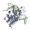
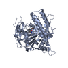
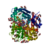
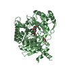
| Title | Structural insights into Pseudomonas aeruginosa Type six secretion system exported effector 8: Tse8 in complex with a peptide | ||||||
 Components Components |
| ||||||
 Keywords Keywords | TOXIN / Amidase Signature (AS) superfamily complex | ||||||
| Function / homology | Amidase signature domain / Amidase signature (AS) superfamily / Amidase / metal ion binding / Amidase domain-containing protein Function and homology information Function and homology information | ||||||
| Biological species |  Pseudomonas aeruginosa PAO1 (bacteria) Pseudomonas aeruginosa PAO1 (bacteria) | ||||||
| Method |  X-RAY DIFFRACTION / SYNCHROTRON / MOLECULAR REPLACEMENT / Resolution: 2.29 Å X-RAY DIFFRACTION / SYNCHROTRON / MOLECULAR REPLACEMENT / Resolution: 2.29 Å | ||||||
 Authors Authors | Sainz-Polo, M.A. / Capuni, R. / Lucas, M. / Altuna, J. / Fucini, P. / Montanchez, I. / Albesa-Jove, D. | ||||||
| Funding support |  Spain, 1items Spain, 1items
| ||||||
 Citation Citation | Journal: J.Struct.Biol. / Year: 2020 Title: Structural insights into Pseudomonas aeruginosaType six secretion system exported effector 8. Authors: Gonzalez-Magana, A. / Sainz-Polo, M.A. / Pretre, G. / Capuni, R. / Lucas, M. / Altuna, J. / Montanchez, I. / Fucini, P. / Albesa-Jove, D. | ||||||
| History |
|
- Structure visualization
| Structure viewer | Molecule: MolmilJmol/JSmol |
|---|
- Downloads & links
-Download
| PDBx/mmCIF format | 6te4.cif.gz | 229.5 KB | Display | PDBx/mmCIF format |
|---|---|---|---|---|
| PDB format | pdb6te4.ent.gz | 180.9 KB | Display | PDB format |
| PDBx/mmJSON format | 6te4.json.gz | Tree view | PDBx/mmJSON format | |
| Others |  Other downloads Other downloads |
-Validation report
| Summary document | 6te4_validation.pdf.gz | 445.4 KB | Display | wwPDB validaton report |
|---|---|---|---|---|
| Full document | 6te4_full_validation.pdf.gz | 450 KB | Display | |
| Data in XML | 6te4_validation.xml.gz | 42.7 KB | Display | |
| Data in CIF | 6te4_validation.cif.gz | 61.6 KB | Display | |
| Arichive directory | https://data.pdbj.org/pub/pdb/validation_reports/te/6te4ftp://data.pdbj.org/pub/pdb/validation_reports/te/6te4 | HTTPS FTP |
-Related structure data
| Related structure data | 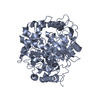 6yhvC 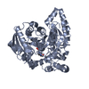 1m22S S: Starting model for refinement C: citing same article ( |
|---|---|
| Similar structure data |


-Links
 PDBj
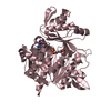
PDBj- Assembly
| Deposited unit | 
| ||||||||||
|---|---|---|---|---|---|---|---|---|---|---|---|
| 1 | 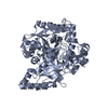
| ||||||||||
| 2 | 
| ||||||||||
| Unit cell |
|
-Components
| #1: Protein | Mass: 63517.316 Da / Num. of mol.: 2 Source method: isolated from a genetically manipulated source Source: (gene. exp.) Pseudomonas aeruginosa PAO1 (bacteria) / Gene: PA4163 / Production host: #2: Protein/peptide | | Mass: 612.738 Da / Num. of mol.: 1 / Source method: isolated from a natural source Details: peptide that might be cleaved from the C-terminal part of the protein as identified by MS analysis. Source: (natural) #3: Water | ChemComp-HOH / |  Mass: 18.015 Da / Num. of mol.: 500 / Source method: isolated from a natural source / Formula: H2O Mass: 18.015 Da / Num. of mol.: 500 / Source method: isolated from a natural source / Formula: H2O |
|---|
-Experimental details
-Experiment
| Experiment | Method: X-RAY DIFFRACTION / Number of used crystals: 1 |
|---|
- Sample preparation
| Crystal | Density Matthews: 2.66 Å3/Da / Density % sol: 53.71 % / Description: prisms |
|---|---|
| Crystal grow | Temperature: 293 K / Method: vapor diffusion, sitting drop / pH: 6.5 Details: 1.6 M magnesium sulfate heptahydrate, 0.1 M MES buffer pH 6.5 |
-Data collection
| Diffraction | Mean temperature: 100 K / Serial crystal experiment: N |
|---|---|
| Diffraction source | Source: SYNCHROTRON / Site: ALBA / Beamline: XALOC / Wavelength: 0.9793 Å |
| Detector | Type: DECTRIS PILATUS3 S 6M / Detector: PIXEL / Date: Mar 17, 2018 |
| Radiation | Protocol: SINGLE WAVELENGTH / Monochromatic (M) / Laue (L): M / Scattering type: x-ray |
| Radiation wavelength | Wavelength: 0.9793 Å / Relative weight: 1 |
| Reflection | Resolution: 2.29→56.632 Å / Num. obs: 53333 / % possible obs: 94.85 % / Redundancy: 3.4 % / Biso Wilson estimate: 22.48 Å2 / CC1/2: 0.979 / CC star: 0.995 / Rmerge(I) obs: 0.1873 / Rpim(I) all: 0.1199 / Rrim(I) all: 0.2233 / Net I/σ(I): 6.96 |
| Reflection shell | Resolution: 2.29→2.32 Å / Redundancy: 3.4 % / Rmerge(I) obs: 0.7321 / Mean I/σ(I) obs: 2.25 / Num. unique obs: 2646 / CC1/2: 0.852 / CC star: 0.959 / R split: 0.4686 / Rrim(I) all: 0.8725 / % possible all: 90.26 |
- Processing
| Software |
| ||||||||||||||||||||||||||||||||||||||||||||||||||||||||||||||||||||||||||||||||||||||||||
|---|---|---|---|---|---|---|---|---|---|---|---|---|---|---|---|---|---|---|---|---|---|---|---|---|---|---|---|---|---|---|---|---|---|---|---|---|---|---|---|---|---|---|---|---|---|---|---|---|---|---|---|---|---|---|---|---|---|---|---|---|---|---|---|---|---|---|---|---|---|---|---|---|---|---|---|---|---|---|---|---|---|---|---|---|---|---|---|---|---|---|---|
| Refinement | Method to determine structure: MOLECULAR REPLACEMENT Starting model: 1m22 Resolution: 2.29→56.632 Å / SU ML: 0.33 / Cross valid method: FREE R-VALUE / σ(F): 1.96 / Phase error: 29.02
| ||||||||||||||||||||||||||||||||||||||||||||||||||||||||||||||||||||||||||||||||||||||||||
| Solvent computation | Shrinkage radii: 0.9 Å / VDW probe radii: 1.11 Å | ||||||||||||||||||||||||||||||||||||||||||||||||||||||||||||||||||||||||||||||||||||||||||
| Displacement parameters | Biso max: 101.97 Å2 / Biso mean: 24.63 Å2 / Biso min: 12.3 Å2 | ||||||||||||||||||||||||||||||||||||||||||||||||||||||||||||||||||||||||||||||||||||||||||
| Refinement step | Cycle: final / Resolution: 2.29→56.632 Å
| ||||||||||||||||||||||||||||||||||||||||||||||||||||||||||||||||||||||||||||||||||||||||||
| Refine LS restraints |
| ||||||||||||||||||||||||||||||||||||||||||||||||||||||||||||||||||||||||||||||||||||||||||
| LS refinement shell | Refine-ID: X-RAY DIFFRACTION / Rfactor Rfree error: 0
|
