 Movie
Movie Controller

[English] 日本語
 Yorodumi
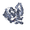
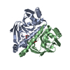
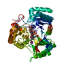
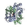
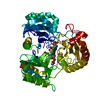
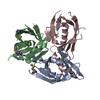
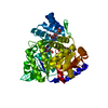
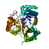
- PDB-1m22: X-ray structure of native peptide amidase from Stenotrophomonas m... -
+ Open data

- Basic information
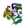
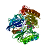
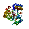
| Entry | Database: PDB / ID: 1m22 | ||||||
|---|---|---|---|---|---|---|---|
| Title | X-ray structure of native peptide amidase from Stenotrophomonas maltophilia at 1.4 A | ||||||
 Components Components | peptide amidase | ||||||
 Keywords Keywords | HYDROLASE / eleven-stranded beta sheet / covered double layers of alpha helices on top and bottom | ||||||
| Function / homology |  Function and homology information Function and homology information | ||||||
| Biological species |  Stenotrophomonas maltophilia (bacteria) Stenotrophomonas maltophilia (bacteria) | ||||||
| Method |  X-RAY DIFFRACTION / SYNCHROTRON / MIRAS / Resolution: 1.4 Å X-RAY DIFFRACTION / SYNCHROTRON / MIRAS / Resolution: 1.4 Å | ||||||
 Authors Authors | Labahn, J. / Neumann, S. / Buldt, G. / Kula, M.-R. / Granzin, J. | ||||||
 Citation Citation | Journal: J.MOL.BIOL. / Year: 2002 Title: An alternative mechanism for amidase signature enzymes Authors: Labahn, J. / Neumann, S. / Buldt, G. / Kula, M.-R. / Granzin, J. #1: Journal: Acta Crystallogr.,Sect.D / Year: 2002Title: Crystallization and preliminary X-ray data of the recombinant peptide amidase from Stenotrophomonas maltophilia Authors: Neumann, S. / Granzin, J. / Kula, M.-R. / Labahn, J. #2: Journal: Appl.Microbiol.Biotechnol. / Year: 1995Title: Purification and characterisation of a newly screened microbial peptide amidase Authors: Stelkes-Ritter, U. / Wyzgol, K. / Kula, M.-R. | ||||||
| History |
|
- Structure visualization
| Structure viewer | Molecule: MolmilJmol/JSmol |
|---|
- Downloads & links
-Download
| PDBx/mmCIF format | 1m22.cif.gz | 218.6 KB | Display | PDBx/mmCIF format |
|---|---|---|---|---|
| PDB format | pdb1m22.ent.gz | 171.3 KB | Display | PDB format |
| PDBx/mmJSON format | 1m22.json.gz | Tree view | PDBx/mmJSON format | |
| Others |  Other downloads Other downloads |
-Validation report
| Arichive directory | https://data.pdbj.org/pub/pdb/validation_reports/m2/1m22ftp://data.pdbj.org/pub/pdb/validation_reports/m2/1m22 | HTTPS FTP |
|---|
-Related structure data
-Links
 PDBj
PDBj


- Assembly
| Deposited unit | 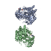
| ||||||||
|---|---|---|---|---|---|---|---|---|---|
| 1 | 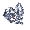
| ||||||||
| 2 | 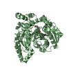
| ||||||||
| Unit cell |
| ||||||||
| Details | the biological unit is the monomer |
-Components
| #1: Protein | Mass: 53545.301 Da / Num. of mol.: 2 Source method: isolated from a genetically manipulated source Source: (gene. exp.) Stenotrophomonas maltophilia (bacteria)Plasmid: pEK06 / Production host: References: UniProt: Q8RJN5, Hydrolases; Acting on carbon-nitrogen bonds, other than peptide bonds; In linear amides #2: Chemical |   Mass: 238.305 Da / Num. of mol.: 2 / Source method: obtained synthetically / Formula: C8H18N2O4S / Comment: pH buffer*YM Mass: 238.305 Da / Num. of mol.: 2 / Source method: obtained synthetically / Formula: C8H18N2O4S / Comment: pH buffer*YM#3: Water | ChemComp-HOH / |  Mass: 18.015 Da / Num. of mol.: 1149 / Source method: isolated from a natural source / Formula: H2O Mass: 18.015 Da / Num. of mol.: 1149 / Source method: isolated from a natural source / Formula: H2O |
|---|
-Experimental details
-Experiment
| Experiment | Method: X-RAY DIFFRACTION / Number of used crystals: 1 |
|---|
- Sample preparation
| Crystal | Density Matthews: 2.21 Å3/Da / Density % sol: 44.29 % | ||||||||||||||||||||||||||||||||||||
|---|---|---|---|---|---|---|---|---|---|---|---|---|---|---|---|---|---|---|---|---|---|---|---|---|---|---|---|---|---|---|---|---|---|---|---|---|---|
| Crystal grow | Temperature: 289 K / Method: vapor diffusion, sitting drop / pH: 7.5 Details: PEG6000, Hepes Glycerine Sodium Azide, pH 7.5, vapour diffusion, sitting drop, temperature 289K | ||||||||||||||||||||||||||||||||||||
| Crystal grow | *PLUS Method: vapor diffusion, sitting dropDetails: Neumann, S., (2002) Acta Crystallogr., Sect.D, 58, 333. | ||||||||||||||||||||||||||||||||||||
| Components of the solutions | *PLUS
|
-Data collection
| Diffraction | Mean temperature: 100 K |
|---|---|
| Diffraction source | Source: SYNCHROTRON / Site: ESRF  / Beamline: ID14-1 / Wavelength: 0.934 Å / Beamline: ID14-1 / Wavelength: 0.934 Å |
| Detector | Type: MARRESEARCH / Detector: CCD / Date: Jul 12, 2001 |
| Radiation | Monochromator: diamond(111), Ge(220) / Protocol: SINGLE WAVELENGTH / Monochromatic (M) / Laue (L): M / Scattering type: x-ray |
| Radiation wavelength | Wavelength: 0.934 Å / Relative weight: 1 |
| Reflection | Resolution: 1.4→79 Å / Num. all: 182206 / Num. obs: 182206 / % possible obs: 99.3 % / Observed criterion σ(F): 0 / Observed criterion σ(I): 0 / Redundancy: 3.9 % / Biso Wilson estimate: 11.9 Å2 / Rmerge(I) obs: 0.103 / Net I/σ(I): 7.8 |
| Reflection shell | Resolution: 1.4→1.49 Å / Rmerge(I) obs: 0.169 / Mean I/σ(I) obs: 5.1 / % possible all: 95 |
| Reflection | *PLUS Lowest resolution: 79 Å / Rmerge(I) obs: 0.103 |
| Reflection shell | *PLUS % possible obs: 95 % / Rmerge(I) obs: 0.169 |
- Processing
| Software |
| ||||||||||||||||||||||||||||||||||||
|---|---|---|---|---|---|---|---|---|---|---|---|---|---|---|---|---|---|---|---|---|---|---|---|---|---|---|---|---|---|---|---|---|---|---|---|---|---|
| Refinement | Method to determine structure: MIRAS / Resolution: 1.4→79 Å / Rfactor Rfree error: 0.002 / Isotropic thermal model: RESTRAINED / Cross valid method: THROUGHOUT / σ(F): 0 / Stereochemistry target values: Engh & Huber
| ||||||||||||||||||||||||||||||||||||
| Solvent computation | Solvent model: FLAT MODEL / Bsol: 44.5075 Å2 / ksol: 0.380289 e/Å3 | ||||||||||||||||||||||||||||||||||||
| Displacement parameters | Biso mean: 15.1 Å2
| ||||||||||||||||||||||||||||||||||||
| Refine analyze |
| ||||||||||||||||||||||||||||||||||||
| Refinement step | Cycle: LAST / Resolution: 1.4→79 Å
| ||||||||||||||||||||||||||||||||||||
| Refine LS restraints |
| ||||||||||||||||||||||||||||||||||||
| Refine LS restraints NCS | NCS model details: CONSTR | ||||||||||||||||||||||||||||||||||||
| LS refinement shell | Resolution: 1.4→1.49 Å / Rfactor Rfree error: 0.007 / Total num. of bins used: 6
| ||||||||||||||||||||||||||||||||||||
| Xplor file |
| ||||||||||||||||||||||||||||||||||||
| Refinement | *PLUS Highest resolution: 1.4 Å / Lowest resolution: 100 Å / Rfactor all: 0.18 / Rfactor obs: 0.189 / Rfactor Rfree: 0.199 / Rfactor Rwork: 0.188 | ||||||||||||||||||||||||||||||||||||
| Solvent computation | *PLUS | ||||||||||||||||||||||||||||||||||||
| Displacement parameters | *PLUS | ||||||||||||||||||||||||||||||||||||
| Refine LS restraints | *PLUS
| ||||||||||||||||||||||||||||||||||||
| LS refinement shell | *PLUS Rfactor Rfree: 0.26 / Rfactor Rwork: 0.25 |
