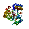
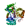
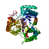
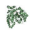
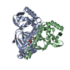
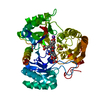
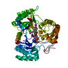
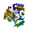
- PDB-3cm1: Crystal structure of SsgA-like sporulation-specific cell division... -
+
Open data
ID or keywords:
Loading...
-
Basic information
Entry
Database: PDB / ID: 3cm1
Title
Crystal structure of SsgA-like sporulation-specific cell division protein (YP_290167.1) from Thermobifida fusca YX-ER1 at 2.60 A resolution
Components
SsgA-like sporulation-specific cell division protein
Keywords
CELL CYCLE / YP_290167.1 / SsgA-like sporulation-specific cell division protein / Streptomyces sporulation and cell division protein / SsgA / Structural Genomics / Joint Center for Structural Genomics / JCSG / Protein Structure Initiative / PSI-2
Function / homology
Function and homology information
positive regulation of FtsZ-dependent cytokinesis / cell septum / division septum assembly / sporulation resulting in formation of a cellular spore Similarity search - Function
Sporulation-specific cell division protein SsgB / Sporulation-specific cell division protein SsgB / Sporulation-specific cell division protein SsgB superfamily / Streptomyces sporulation and cell division protein, SsgA / Transcriptional Co-activator pc4; Chain A / Roll / Mainly Beta Similarity search - Domain/homology
Mass: 15415.895 Da / Num. of mol.: 3 Source method: isolated from a genetically manipulated source Source: (gene. exp.) Thermobifida fusca (bacteria) / Strain: YX / Gene: YP_290167.1, Tfu_2111 / Plasmid: SpeedET / Production host: Escherichia coli (E. coli) / Strain (production host): HK100 / References: UniProt: Q47N25
Has protein modification
Y
Sequence details
THE CONSTRUCT WAS EXPRESSED WITH A PURIFICATION TAG MGSDKIHHHHHHENLYFQG. THE TAG WAS REMOVED WITH ...THE CONSTRUCT WAS EXPRESSED WITH A PURIFICATION TAG MGSDKIHHHHHHENLYFQG. THE TAG WAS REMOVED WITH TEV PROTEASE LEAVING ONLY A GLYCINE (0) FOLLOWED BY THE TARGET SEQUENCE.
-
Experimental details
-
Experiment
Experiment
Method: X-RAY DIFFRACTION / Number of used crystals: 1
-
Sample preparation
Crystal
Density Matthews: 2.97 Å3/Da / Density % sol: 58.56 %
Crystal grow
Temperature: 277 K / Method: vapor diffusion, sitting drop / pH: 4.5 Details: NANODROP, 40.0% 1,2-propanediol, 0.1M Acetate pH 4.5, VAPOR DIFFUSION, SITTING DROP, temperature 277K
Type: MARMOSAIC 325 mm CCD / Detector: CCD / Date: May 13, 2007 / Details: Flat mirror (vertical focusing)
Radiation
Monochromator: Single crystal Si(111) bent (horizontal focusing) Protocol: MAD / Monochromatic (M) / Laue (L): M / Scattering type: x-ray
Radiation wavelength
ID
Wavelength (Å)
Relative weight
1
0.91837
1
2
0.97929
1
3
0.97898
1
Reflection
Resolution: 2.6→46.029 Å / Num. obs: 16529 / % possible obs: 99.4 % / Observed criterion σ(I): -3 / Biso Wilson estimate: 79.297 Å2 / Rmerge(I) obs: 0.046 / Net I/σ(I): 15.84
Reflection shell
Resolution (Å)
Rmerge(I) obs
Mean I/σ(I) obs
Num. measured obs
Num. unique obs
Diffraction-ID
% possible all
2.6-2.69
0.793
1.8
5930
1579
1
98.9
2.69-2.8
0.554
2.7
6435
1683
1
99.9
2.8-2.93
0.373
3.9
6450
1689
1
99.9
2.93-3.08
0.244
5.7
6203
1625
1
99.9
3.08-3.27
0.146
8.6
6196
1621
1
99.9
3.27-3.52
0.082
13.9
6303
1646
1
100
3.52-3.88
0.049
20.5
6493
1698
1
99.8
3.88-4.43
0.031
28.9
6255
1637
1
99.8
4.43-5.57
0.026
35
6369
1683
1
99.9
5.57-46.029
0.027
36.5
5934
1666
1
96.4
-
Phasing
Phasing
Method: MAD
-
Processing
Software
Name
Version
Classification
NB
REFMAC
5.2.0019
refinement
PHENIX
refinement
SHELX
phasing
MolProbity
3beta29
modelbuilding
XSCALE
datascaling
PDB_EXTRACT
3
dataextraction
MAR345
CCD
datacollection
XDS
datareduction
SHELXD
phasing
autoSHARP
phasing
Refinement
Method to determine structure: MAD / Resolution: 2.6→46.029 Å / Cor.coef. Fo:Fc: 0.937 / Cor.coef. Fo:Fc free: 0.915 / SU B: 29.904 / SU ML: 0.277 / TLS residual ADP flag: LIKELY RESIDUAL / Cross valid method: THROUGHOUT / σ(F): 0 / ESU R: 0.513 / ESU R Free: 0.308 Stereochemistry target values: MAXIMUM LIKELIHOOD WITH PHASES Details: 1. HYDROGENS HAVE BEEN ADDED IN THE RIDING POSITIONS. 2. A MET-INHIBITION PROTOCOL WAS USED FOR SELENOMETHIONINE INCORPORATION DURING PROTEIN EXPRESSION. THE OCCUPANCY OF THE SE ATOMS IN THE ...Details: 1. HYDROGENS HAVE BEEN ADDED IN THE RIDING POSITIONS. 2. A MET-INHIBITION PROTOCOL WAS USED FOR SELENOMETHIONINE INCORPORATION DURING PROTEIN EXPRESSION. THE OCCUPANCY OF THE SE ATOMS IN THE MSE RESIDUES WAS REDUCED TO 0.75 FOR THE REDUCED SCATTERING POWER DUE TO PARTIAL S-MET INCORPORATION. 3. ATOM RECORD CONTAINS RESIDUAL B FACTORS ONLY.
Rfactor
Num. reflection
% reflection
Selection details
Rfree
0.27
846
5.1 %
RANDOM
Rwork
0.23
-
-
-
obs
0.232
16493
99.44 %
-
Solvent computation
Ion probe radii: 0.8 Å / Shrinkage radii: 0.8 Å / VDW probe radii: 1.2 Å / Solvent model: BABINET MODEL WITH MASK
Displacement parameters
Biso mean: 63.264 Å2
Baniso -1
Baniso -2
Baniso -3
1-
2.08 Å2
0 Å2
0 Å2
2-
-
2.08 Å2
0 Å2
3-
-
-
-4.15 Å2
Refinement step
Cycle: LAST / Resolution: 2.6→46.029 Å
Protein
Nucleic acid
Ligand
Solvent
Total
Num. atoms
2892
0
0
0
2892
Refine LS restraints
Refine-ID
Type
Dev ideal
Dev ideal target
Number
X-RAY DIFFRACTION
r_bond_refined_d
0.011
0.022
2952
X-RAY DIFFRACTION
r_bond_other_d
0.002
0.02
1905
X-RAY DIFFRACTION
r_angle_refined_deg
1.414
1.952
4023
X-RAY DIFFRACTION
r_angle_other_deg
1.012
3
4634
X-RAY DIFFRACTION
r_dihedral_angle_1_deg
4.67
5
373
X-RAY DIFFRACTION
r_dihedral_angle_2_deg
29.738
23.75
128
X-RAY DIFFRACTION
r_dihedral_angle_3_deg
16.165
15
425
X-RAY DIFFRACTION
r_dihedral_angle_4_deg
21.95
15
20
X-RAY DIFFRACTION
r_chiral_restr
0.092
0.2
465
X-RAY DIFFRACTION
r_gen_planes_refined
0.005
0.02
3307
X-RAY DIFFRACTION
r_gen_planes_other
0.002
0.02
590
X-RAY DIFFRACTION
r_nbd_refined
0.206
0.2
537
X-RAY DIFFRACTION
r_nbd_other
0.175
0.2
1759
X-RAY DIFFRACTION
r_nbtor_refined
0.179
0.2
1396
X-RAY DIFFRACTION
r_nbtor_other
0.087
0.2
1620
X-RAY DIFFRACTION
r_xyhbond_nbd_refined
0.127
0.2
38
X-RAY DIFFRACTION
r_symmetry_vdw_refined
0.182
0.2
4
X-RAY DIFFRACTION
r_symmetry_vdw_other
0.248
0.2
20
X-RAY DIFFRACTION
r_symmetry_hbond_refined
0.07
0.2
2
X-RAY DIFFRACTION
r_mcbond_it
1.396
3
1941
X-RAY DIFFRACTION
r_mcbond_other
0.203
3
765
X-RAY DIFFRACTION
r_mcangle_it
2.408
5
3044
X-RAY DIFFRACTION
r_scbond_it
4.446
8
1136
X-RAY DIFFRACTION
r_scangle_it
6.667
11
979
Refine LS restraints NCS
Ens-ID: 1 / Refine-ID: X-RAY DIFFRACTION
Dom-ID
Auth asym-ID
Number
Type
Rms dev position (Å)
Weight position
1
A
595
TIGHTPOSITIONAL
0.06
0.05
2
B
595
TIGHTPOSITIONAL
0.05
0.05
3
C
595
TIGHTPOSITIONAL
0.05
0.05
1
A
655
MEDIUMPOSITIONAL
0.35
0.25
2
B
655
MEDIUMPOSITIONAL
0.34
0.25
3
C
655
MEDIUMPOSITIONAL
0.29
0.25
1
A
595
TIGHTTHERMAL
0.09
0.5
2
B
595
TIGHTTHERMAL
0.07
0.5
3
C
595
TIGHTTHERMAL
0.08
0.5
1
A
655
MEDIUMTHERMAL
0.25
1
2
B
655
MEDIUMTHERMAL
0.23
1
3
C
655
MEDIUMTHERMAL
0.23
1
LS refinement shell
Resolution: 2.6→2.668 Å / Total num. of bins used: 20
Rfactor
Num. reflection
% reflection
Rfree
0.432
81
-
Rwork
0.383
1115
-
all
-
1196
-
obs
-
-
98.52 %
Refinement TLS params.
Method: refined / Refine-ID: X-RAY DIFFRACTION
ID
L11 (°2)
L12 (°2)
L13 (°2)
L22 (°2)
L23 (°2)
L33 (°2)
S11 (Å °)
S12 (Å °)
S13 (Å °)
S21 (Å °)
S22 (Å °)
S23 (Å °)
S31 (Å °)
S32 (Å °)
S33 (Å °)
T11 (Å2)
T12 (Å2)
T13 (Å2)
T22 (Å2)
T23 (Å2)
T33 (Å2)
Origin x (Å)
Origin y (Å)
Origin z (Å)
1
3.3237
-0.7608
1.4573
3.1621
0.7292
4.4989
-0.2266
-0.3363
0.0151
0.4539
0.0638
-0.1049
0.1261
-0.2993
0.1628
-0.2305
-0.0629
0.0873
-0.0691
-0.0743
-0.1591
24.7602
20.6761
-4.0513
2
2.2034
0.6055
0.9095
5.7829
0.207
4.3504
0.1708
-0.3358
-0.4891
0.4645
-0.0447
0.5138
0.64
-0.3466
-0.1261
0.263
-0.1129
0.1789
-0.0825
-0.023
0.1467
18.8947
-2.4831
-11.6394
3
3.0642
0.186
0.7971
5.3186
0.0636
3.9722
0.031
0.1665
-0.0475
-0.3227
-0.1135
0.9923
0.1933
-0.4303
0.0825
-0.1477
-0.1014
0.036
-0.0849
-0.0739
0.0501
12.0823
17.0616
-23.8404
Refinement TLS group
ID
Refine-ID
Refine TLS-ID
Auth asym-ID
Label asym-ID
Auth seq-ID
Label seq-ID
1
X-RAY DIFFRACTION
1
A
A
1 - 137
2 - 138
2
X-RAY DIFFRACTION
2
B
B
1 - 137
2 - 138
3
X-RAY DIFFRACTION
3
C
C
1 - 137
2 - 138
+
About Yorodumi
-
News
-
Feb 9, 2022. New format data for meta-information of EMDB entries
New format data for meta-information of EMDB entries
Version 3 of the EMDB header file is now the official format.
The previous official version 1.9 will be removed from the archive.
In the structure databanks used in Yorodumi, some data are registered as the other names, "COVID-19 virus" and "2019-nCoV". Here are the details of the virus and the list of structure data.
Jan 31, 2019. EMDB accession codes are about to change! (news from PDBe EMDB page)
EMDB accession codes are about to change! (news from PDBe EMDB page)
The allocation of 4 digits for EMDB accession codes will soon come to an end. Whilst these codes will remain in use, new EMDB accession codes will include an additional digit and will expand incrementally as the available range of codes is exhausted. The current 4-digit format prefixed with “EMD-” (i.e. EMD-XXXX) will advance to a 5-digit format (i.e. EMD-XXXXX), and so on. It is currently estimated that the 4-digit codes will be depleted around Spring 2019, at which point the 5-digit format will come into force.
The EM Navigator/Yorodumi systems omit the EMD- prefix.
Related info.:Q: What is EMD? / ID/Accession-code notation in Yorodumi/EM Navigator
Yorodumi is a browser for structure data from EMDB, PDB, SASBDB, etc.
This page is also the successor to EM Navigator detail page, and also detail information page/front-end page for Omokage search.
The word "yorodu" (or yorozu) is an old Japanese word meaning "ten thousand". "mi" (miru) is to see.
Related info.:EMDB / PDB / SASBDB / Comparison of 3 databanks / Yorodumi Search / Aug 31, 2016. New EM Navigator & Yorodumi / Yorodumi Papers / Jmol/JSmol / Function and homology information / Changes in new EM Navigator and Yorodumi
 Movie
Movie Controller

 Yorodumi
 Open data
 Basic information
 Components
 Keywords
 Function and homology information
 Thermobifida fusca (bacteria)
 X-RAY DIFFRACTION /
  Authors
 Citation
 Structure visualization
 Downloads & links
 Other downloads
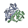

 PDBj
PDBj Assembly
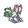
 Sample preparation
 / Beamline: BL11-1 / Wavelength: 0.91837, 0.97929, 0.97898
 Processing