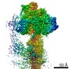
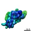
 Movie
Movie Controller

[English] 日本語
 Yorodumi
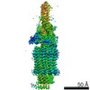
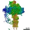
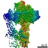
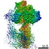
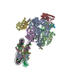
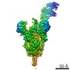
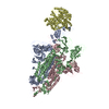
- PDB-6te0: Cryo-EM structure of Euglena gracilis mitochondrial ATP synthase,... -
+ Open data

- Basic information
| Entry | Database: PDB / ID: 6te0 | ||||||
|---|---|---|---|---|---|---|---|
| Title | Cryo-EM structure of Euglena gracilis mitochondrial ATP synthase, OSCP/F1/c-ring, rotational state 3 | ||||||
 Components Components |
| ||||||
 Keywords Keywords | MEMBRANE PROTEIN / mitochondria / ATP synthase | ||||||
| Function / homology |  Function and homology information Function and homology informationThrombin, subunit H - #170 / F1F0 ATP synthase subunit C / F1FO ATP Synthase / Bovine Mitochondrial F1-ATPase, ATP Synthase Beta Chain; Chain D, domain3 / Bovine Mitochondrial F1-atpase; Atp Synthase Beta Chain; Chain D, domain 3 / Thrombin, subunit H / P-loop containing nucleotide triphosphate hydrolases / Up-down Bundle / Beta Barrel / Rossmann fold ...Thrombin, subunit H - #170 / F1F0 ATP synthase subunit C / F1FO ATP Synthase / Bovine Mitochondrial F1-ATPase, ATP Synthase Beta Chain; Chain D, domain3 / Bovine Mitochondrial F1-atpase; Atp Synthase Beta Chain; Chain D, domain 3 / Thrombin, subunit H / P-loop containing nucleotide triphosphate hydrolases / Up-down Bundle / Beta Barrel / Rossmann fold / Orthogonal Bundle / 3-Layer(aba) Sandwich / Mainly Beta / Mainly Alpha / Alpha Beta Similarity search - Domain/homology | ||||||
| Biological species |  Euglena gracilis (euglena) Euglena gracilis (euglena) | ||||||
| Method | ELECTRON MICROSCOPY / single particle reconstruction / cryo EM / Resolution: 3.92 Å | ||||||
 Authors Authors | Muhleip, A. / Amunts, A. | ||||||
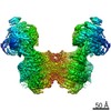
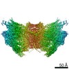
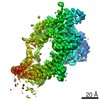
 Citation Citation | Journal: Elife / Year: 2019 Title: Structure of a mitochondrial ATP synthase with bound native cardiolipin. Authors: Alexander Mühleip / Sarah E McComas / Alexey Amunts /  Abstract: The mitochondrial ATP synthase fuels eukaryotic cells with chemical energy. Here we report the cryo-EM structure of a divergent ATP synthase dimer from mitochondria of , a member of the phylum ...The mitochondrial ATP synthase fuels eukaryotic cells with chemical energy. Here we report the cryo-EM structure of a divergent ATP synthase dimer from mitochondria of , a member of the phylum Euglenozoa that also includes human parasites. It features 29 different subunits, 8 of which are newly identified. The membrane region was determined to 2.8 Å resolution, enabling the identification of 37 associated lipids, including 25 cardiolipins, which provides insight into protein-lipid interactions and their functional roles. The rotor-stator interface comprises four membrane-embedded horizontal helices, including a distinct subunit . The dimer interface is formed entirely by phylum-specific components, and a peripherally associated subcomplex contributes to the membrane curvature. The central and peripheral stalks directly interact with each other. Last, the ATPase inhibitory factor 1 (IF) binds in a mode that is different from human, but conserved in Trypanosomatids. | ||||||
| History |
|
- Structure visualization
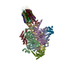
| Movie |
Movie viewer |
|---|---|
| Structure viewer | Molecule: MolmilJmol/JSmol |
- Downloads & links
-Download
| PDBx/mmCIF format | 6te0.cif.gz | 1.6 MB | Display | PDBx/mmCIF format |
|---|---|---|---|---|
| PDB format | pdb6te0.ent.gz | 1.3 MB | Display | PDB format |
| PDBx/mmJSON format | 6te0.json.gz | Tree view | PDBx/mmJSON format | |
| Others |  Other downloads Other downloads |
-Validation report
| Arichive directory | https://data.pdbj.org/pub/pdb/validation_reports/te/6te0ftp://data.pdbj.org/pub/pdb/validation_reports/te/6te0 | HTTPS FTP |
|---|
-Related structure data
| Related structure data | 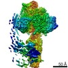 10473MC  6tduC  6tdvC 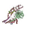 6tdwC  6tdxC  6tdyC 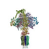 6tdzC M: map data used to model this data C: citing same article ( |
|---|---|
| Similar structure data |


-Links
 PDBj
PDBj
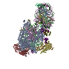
- Assembly
| Deposited unit | 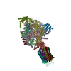
|
|---|---|
| 1 |
|
-Components
-ATP synthase subunit ... , 5 types, 18 molecules CABFDEHIOPQRSTUVWX
| #1: Protein | Mass: 61897.199 Da / Num. of mol.: 3 / Source method: isolated from a natural source / Source: (natural) Euglena gracilis (euglena)#2: Protein | Mass: 53213.035 Da / Num. of mol.: 3 / Source method: isolated from a natural source / Source: (natural) Euglena gracilis (euglena)#6: Protein | | Mass: 19559.953 Da / Num. of mol.: 1 / Source method: isolated from a natural source / Source: (natural) Euglena gracilis (euglena)#7: Protein | | Mass: 8751.981 Da / Num. of mol.: 1 / Source method: isolated from a natural source / Source: (natural) Euglena gracilis (euglena)#8: Protein | Mass: 10820.831 Da / Num. of mol.: 10 / Source method: isolated from a natural source / Source: (natural) Euglena gracilis (euglena) |
|---|
-Protein , 3 types, 5 molecules LJKMG
| #3: Protein | Mass: 20995.094 Da / Num. of mol.: 3 / Source method: isolated from a natural source / Source: (natural) Euglena gracilis (euglena)#4: Protein | | Mass: 29564.520 Da / Num. of mol.: 1 / Source method: isolated from a natural source / Source: (natural) Euglena gracilis (euglena)#5: Protein | | Mass: 35110.375 Da / Num. of mol.: 1 / Source method: isolated from a natural source / Source: (natural) Euglena gracilis (euglena) |
|---|
-Non-polymers , 4 types, 11 molecules 


| #9: Chemical | ChemComp-ATP /  Mass: 507.181 Da / Num. of mol.: 4 / Source method: obtained synthetically / Formula: C10H16N5O13P3 / Feature type: SUBJECT OF INVESTIGATION / Comment: ATP, energy-carrying molecule*YM Mass: 507.181 Da / Num. of mol.: 4 / Source method: obtained synthetically / Formula: C10H16N5O13P3 / Feature type: SUBJECT OF INVESTIGATION / Comment: ATP, energy-carrying molecule*YM#10: Chemical | ChemComp-MG /  Mass: 24.305 Da / Num. of mol.: 5 / Source method: obtained synthetically / Formula: Mg Mass: 24.305 Da / Num. of mol.: 5 / Source method: obtained synthetically / Formula: Mg#11: Chemical | ChemComp-ADP / |  Mass: 427.201 Da / Num. of mol.: 1 / Source method: obtained synthetically / Formula: C10H15N5O10P2 / Feature type: SUBJECT OF INVESTIGATION / Comment: ADP, energy-carrying molecule*YM Mass: 427.201 Da / Num. of mol.: 1 / Source method: obtained synthetically / Formula: C10H15N5O10P2 / Feature type: SUBJECT OF INVESTIGATION / Comment: ADP, energy-carrying molecule*YM#12: Chemical | ChemComp-TRT / |  Mass: 352.508 Da / Num. of mol.: 1 / Source method: obtained synthetically / Formula: C21H36O4 / Comment: detergent*YM Mass: 352.508 Da / Num. of mol.: 1 / Source method: obtained synthetically / Formula: C21H36O4 / Comment: detergent*YM |
|---|
-Details
| Has ligand of interest | Y |
|---|
-Experimental details
-Experiment
| Experiment | Method: ELECTRON MICROSCOPY |
|---|---|
| EM experiment | Aggregation state: PARTICLE / 3D reconstruction method: single particle reconstruction |
- Sample preparation
| Component | Name: mitochondrial ATP synthase dimer of Euglena gracilis / Type: COMPLEX / Entity ID: #1-#8 / Source: NATURAL |
|---|---|
| Molecular weight | Value: 2 MDa / Experimental value: NO |
| Source (natural) | Organism: Euglena gracilis (euglena) |
| Buffer solution | pH: 7.4 |
| Specimen | Embedding applied: NO / Shadowing applied: NO / Staining applied: NO / Vitrification applied: YES |
| Vitrification | Instrument: FEI VITROBOT MARK IV / Cryogen name: ETHANE / Humidity: 100 % / Chamber temperature: 277 K / Details: 5 seconds blot |
- Electron microscopy imaging
| Experimental equipment |  Model: Titan Krios / Image courtesy: FEI Company |
|---|---|
| Microscopy | Model: FEI TITAN KRIOS |
| Electron gun | Electron source:  FIELD EMISSION GUN / Accelerating voltage: 300 kV / Illumination mode: FLOOD BEAM FIELD EMISSION GUN / Accelerating voltage: 300 kV / Illumination mode: FLOOD BEAM |
| Electron lens | Mode: BRIGHT FIELD / Nominal magnification: 130000 X / Cs: 2.7 mm / C2 aperture diameter: 70 µm / Alignment procedure: ZEMLIN TABLEAU |
| Specimen holder | Cryogen: NITROGEN / Specimen holder model: FEI TITAN KRIOS AUTOGRID HOLDER |
| Image recording | Average exposure time: 10 sec. / Electron dose: 36.3 e/Å2 / Detector mode: COUNTING / Film or detector model: GATAN K2 QUANTUM (4k x 4k) / Num. of real images: 9045 |
| EM imaging optics | Energyfilter name: GIF Quantum LS / Energyfilter slit width: 20 eV |
- Processing
| EM software |
| |||||||||||||||||||||
|---|---|---|---|---|---|---|---|---|---|---|---|---|---|---|---|---|---|---|---|---|---|---|
| CTF correction | Type: PHASE FLIPPING AND AMPLITUDE CORRECTION | |||||||||||||||||||||
| Particle selection | Num. of particles selected: 555269 | |||||||||||||||||||||
| Symmetry | Point symmetry: C1 (asymmetric) | |||||||||||||||||||||
| 3D reconstruction | Resolution: 3.92 Å / Resolution method: FSC 0.143 CUT-OFF / Num. of particles: 43232 / Algorithm: FOURIER SPACE / Symmetry type: POINT |
