Movie
Movie Controller

[English] 日本語
 Yorodumi
- EMDB-10473: Cryo-EM structure of Euglena gracilis mitochondrial ATP synthase,... -
+ Open data

- Basic information
| Entry | Database: EMDB / ID: EMD-10473 | |||||||||
|---|---|---|---|---|---|---|---|---|---|---|
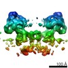
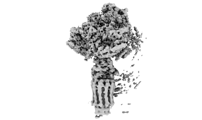
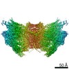
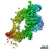
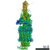
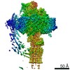
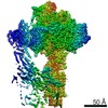
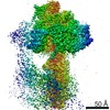
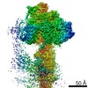
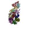
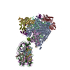
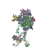
| Title | Cryo-EM structure of Euglena gracilis mitochondrial ATP synthase, OSCP/F1/c-ring, rotational state 3 | |||||||||
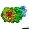
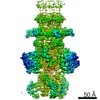
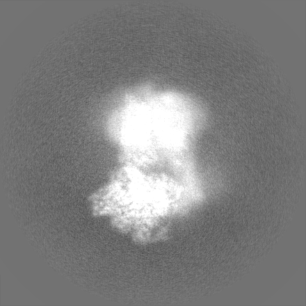
 Map data Map data | local resolution filtered map | |||||||||
 Sample Sample |
| |||||||||
 Keywords Keywords | mitochondria / ATP synthase / MEMBRANE PROTEIN | |||||||||
| Biological species |  Euglena gracilis (euglena) Euglena gracilis (euglena) | |||||||||
| Method | single particle reconstruction / cryo EM / Resolution: 3.92 Å | |||||||||
 Authors Authors | Muhleip A / Amunts A | |||||||||
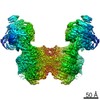
 Citation Citation | Journal: Elife / Year: 2019 Title: Structure of a mitochondrial ATP synthase with bound native cardiolipin. Authors: Alexander Mühleip / Sarah E McComas / Alexey Amunts /  Abstract: The mitochondrial ATP synthase fuels eukaryotic cells with chemical energy. Here we report the cryo-EM structure of a divergent ATP synthase dimer from mitochondria of , a member of the phylum ...The mitochondrial ATP synthase fuels eukaryotic cells with chemical energy. Here we report the cryo-EM structure of a divergent ATP synthase dimer from mitochondria of , a member of the phylum Euglenozoa that also includes human parasites. It features 29 different subunits, 8 of which are newly identified. The membrane region was determined to 2.8 Å resolution, enabling the identification of 37 associated lipids, including 25 cardiolipins, which provides insight into protein-lipid interactions and their functional roles. The rotor-stator interface comprises four membrane-embedded horizontal helices, including a distinct subunit . The dimer interface is formed entirely by phylum-specific components, and a peripherally associated subcomplex contributes to the membrane curvature. The central and peripheral stalks directly interact with each other. Last, the ATPase inhibitory factor 1 (IF) binds in a mode that is different from human, but conserved in Trypanosomatids. | |||||||||
| History |
|
- Structure visualization
| Movie |
 Movie viewer Movie viewer |
|---|---|
| Structure viewer | EM map: SurfViewMolmilJmol/JSmol |
| Supplemental images |
- Downloads & links
-EMDB archive
| Map data | emd_10473.map.gz | 186.2 MB | EMDB map data format | |
|---|---|---|---|---|
| Header (meta data) | emd-10473-v30.xmlemd-10473.xml | 23.3 KB 23.3 KB | Display Display | EMDB header |
| Images |  emd_10473.png emd_10473.png | 53 KB | ||
| Masks | emd_10473_msk_1.map | 325 MB | Mask map | |
| Filedesc metadata | emd-10473.cif.gz | 7.1 KB | ||
| Others | emd_10473_half_map_1.map.gzemd_10473_half_map_2.map.gz | 259.8 MB 259.7 MB | ||
| Archive directory |  http://ftp.pdbj.org/pub/emdb/structures/EMD-10473ftp://ftp.pdbj.org/pub/emdb/structures/EMD-10473 http://ftp.pdbj.org/pub/emdb/structures/EMD-10473ftp://ftp.pdbj.org/pub/emdb/structures/EMD-10473 | HTTPS FTP |
-Related structure data
| Related structure data | 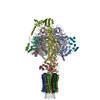 6te0MC  6tduC  6tdvC 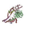 6tdwC 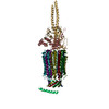 6tdxC 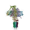 6tdyC 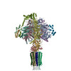 6tdzC M: atomic model generated by this map C: citing same article ( |
|---|---|
| Similar structure data |
-Links
| EMDB pages | EMDB (EBI/PDBe) / EMDataResource |
|---|
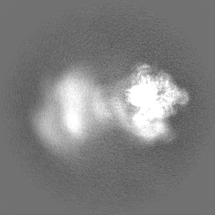
-Map
| File | Download / File: emd_10473.map.gz / Format: CCP4 / Size: 325 MB / Type: IMAGE STORED AS FLOATING POINT NUMBER (4 BYTES) | ||||||||||||||||||||||||||||||||||||||||||||||||||||||||||||||||||||
|---|---|---|---|---|---|---|---|---|---|---|---|---|---|---|---|---|---|---|---|---|---|---|---|---|---|---|---|---|---|---|---|---|---|---|---|---|---|---|---|---|---|---|---|---|---|---|---|---|---|---|---|---|---|---|---|---|---|---|---|---|---|---|---|---|---|---|---|---|---|
| Annotation | local resolution filtered map | ||||||||||||||||||||||||||||||||||||||||||||||||||||||||||||||||||||
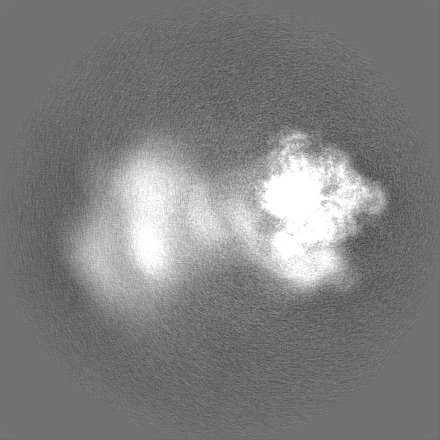
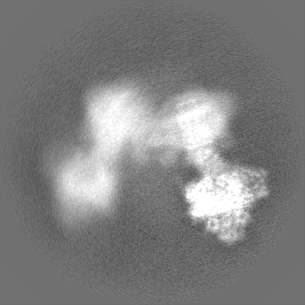
| Projections & slices | Image control
Images are generated by Spider. | ||||||||||||||||||||||||||||||||||||||||||||||||||||||||||||||||||||
| Voxel size | X=Y=Z: 1.05 Å | ||||||||||||||||||||||||||||||||||||||||||||||||||||||||||||||||||||
| Density |
| ||||||||||||||||||||||||||||||||||||||||||||||||||||||||||||||||||||
| Symmetry | Space group: 1 | ||||||||||||||||||||||||||||||||||||||||||||||||||||||||||||||||||||
| Details | EMDB XML:
CCP4 map header:
| ||||||||||||||||||||||||||||||||||||||||||||||||||||||||||||||||||||
 Z (Sec.)
 Y (Row.)
 X (Col.)


-Supplemental data
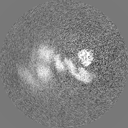
-Mask #1
| File | emd_10473_msk_1.map | ||||||||||||
|---|---|---|---|---|---|---|---|---|---|---|---|---|---|
| Projections & Slices |
| ||||||||||||
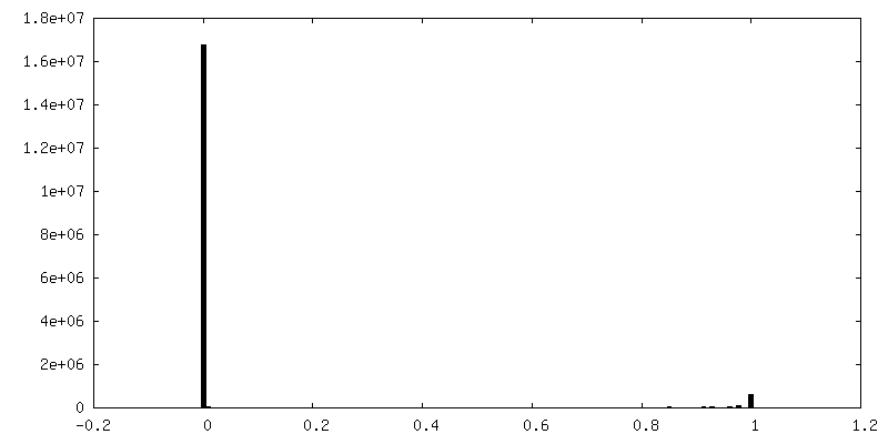
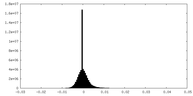
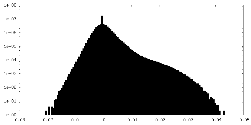
| Density Histograms |


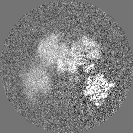
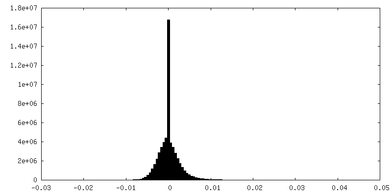
-Half map: #1
| File | emd_10473_half_map_1.map | ||||||||||||
|---|---|---|---|---|---|---|---|---|---|---|---|---|---|
| Projections & Slices |
| ||||||||||||
| Density Histograms |

-Half map: #2
| File | emd_10473_half_map_2.map | ||||||||||||
|---|---|---|---|---|---|---|---|---|---|---|---|---|---|
| Projections & Slices |
| ||||||||||||
| Density Histograms |


- Sample components
+Entire : mitochondrial ATP synthase dimer of Euglena gracilis
+Supramolecule #1: mitochondrial ATP synthase dimer of Euglena gracilis
+Macromolecule #1: ATP synthase subunit alpha
+Macromolecule #2: ATP synthase subunit beta
+Macromolecule #3: Ribonucleoprotein P18
+Macromolecule #4: oligomycin sensitivity conferring protein (OSCP)
+Macromolecule #5: ATP synthase F1 subunit gamma protein
+Macromolecule #6: ATP synthase subunit delta
+Macromolecule #7: ATP synthase subunit epsilon
+Macromolecule #8: ATP synthase subunit c
+Macromolecule #9: ADENOSINE-5'-TRIPHOSPHATE

+Macromolecule #10: MAGNESIUM ION
+Macromolecule #11: ADENOSINE-5'-DIPHOSPHATE

+Macromolecule #12: FRAGMENT OF TRITON X-100

-Experimental details
-Structure determination
| Method | cryo EM |
|---|---|
 Processing Processing | single particle reconstruction |
| Aggregation state | particle |
-Sample preparation
| Buffer | pH: 7.4 |
|---|---|
| Vitrification | Cryogen name: ETHANE / Chamber humidity: 100 % / Chamber temperature: 277 K / Instrument: FEI VITROBOT MARK IV / Details: 5 seconds blot. |
- Electron microscopy
| Microscope | FEI TITAN KRIOS |
|---|---|
| Specialist optics | Energy filter - Name: GIF Quantum LS / Energy filter - Slit width: 20 eV |
| Image recording | Film or detector model: GATAN K2 QUANTUM (4k x 4k) / Detector mode: COUNTING / Number real images: 9045 / Average exposure time: 10.0 sec. / Average electron dose: 36.3 e/Å2 |
| Electron beam | Acceleration voltage: 300 kV / Electron source:  FIELD EMISSION GUN FIELD EMISSION GUN |
| Electron optics | C2 aperture diameter: 70.0 µm / Illumination mode: FLOOD BEAM / Imaging mode: BRIGHT FIELD / Cs: 2.7 mm / Nominal magnification: 130000 |
| Sample stage | Specimen holder model: FEI TITAN KRIOS AUTOGRID HOLDER / Cooling holder cryogen: NITROGEN |
| Experimental equipment |  Model: Titan Krios / Image courtesy: FEI Company |