+ Open data
- Basic information
| Entry | Database: PDB / ID: 6tax | ||||||
|---|---|---|---|---|---|---|---|
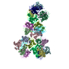
| Title | Mouse RNF213 wild type protein | ||||||
 Components Components | RNF213,E3 ubiquitin-protein ligase RNF213,E3 ubiquitin-protein ligase RNF213 | ||||||
 Keywords Keywords | SIGNALING PROTEIN / RNF213 / E3 LIgase / AAA-Protein | ||||||
| Function / homology |  Function and homology information Function and homology informationlipid ubiquitination / negative regulation of non-canonical Wnt signaling pathway / lipid droplet formation / xenophagy / Antigen processing: Ubiquitination & Proteasome degradation / Transferases; Acyltransferases; Aminoacyltransferases / sprouting angiogenesis / immune system process / protein K63-linked ubiquitination / regulation of lipid metabolic process ...lipid ubiquitination / negative regulation of non-canonical Wnt signaling pathway / lipid droplet formation / xenophagy / Antigen processing: Ubiquitination & Proteasome degradation / Transferases; Acyltransferases; Aminoacyltransferases / sprouting angiogenesis / immune system process / protein K63-linked ubiquitination / regulation of lipid metabolic process / protein autoubiquitination / lipid droplet / Hydrolases; Acting on acid anhydrides; Acting on acid anhydrides to facilitate cellular and subcellular movement / RING-type E3 ubiquitin transferase / ubiquitin-protein transferase activity / ubiquitin protein ligase activity / ubiquitin-dependent protein catabolic process / angiogenesis / defense response to bacterium / protein ubiquitination / nucleolus / ATP hydrolysis activity / ATP binding / metal ion binding / cytosol / cytoplasm Similarity search - Function | ||||||
| Biological species |  | ||||||
| Method | ELECTRON MICROSCOPY / single particle reconstruction / cryo EM / Resolution: 3.2 Å | ||||||
 Authors Authors | Ahel, J. / Meinhart, A. / Haselbach, D. / Clausen, T. | ||||||
| Funding support |  Austria, 1items Austria, 1items
| ||||||
 Citation Citation |  Journal: Elife / Year: 2020 Journal: Elife / Year: 2020Title: Moyamoya disease factor RNF213 is a giant E3 ligase with a dynein-like core and a distinct ubiquitin-transfer mechanism. Authors: Juraj Ahel / Anita Lehner / Antonia Vogel / Alexander Schleiffer / Anton Meinhart / David Haselbach / Tim Clausen /  Abstract: RNF213 is the major susceptibility factor for Moyamoya disease, a progressive cerebrovascular disorder that often leads to brain stroke in adults and children. Characterization of disease-associated ...RNF213 is the major susceptibility factor for Moyamoya disease, a progressive cerebrovascular disorder that often leads to brain stroke in adults and children. Characterization of disease-associated mutations has been complicated by the enormous size of RNF213. Here, we present the cryo-EM structure of mouse RNF213. The structure reveals the intricate fold of the 584 kDa protein, comprising an N-terminal stalk, a dynein-like core with six ATPase units, and a multidomain E3 module. Collaboration with UbcH7, a cysteine-reactive E2, points to an unexplored ubiquitin-transfer mechanism that proceeds in a RING-independent manner. Moreover, we show that pathologic MMD mutations cluster in the composite E3 domain, likely interfering with substrate ubiquitination. In conclusion, the structure of RNF213 uncovers a distinct type of an E3 enzyme, highlighting the growing mechanistic diversity in ubiquitination cascades. Our results also provide the molecular framework for investigating the emerging role of RNF213 in lipid metabolism, hypoxia, and angiogenesis. | ||||||
| History |
|
- Structure visualization
| Movie |
 Movie viewer Movie viewer |
|---|---|
| Structure viewer | Molecule:  Molmil Molmil Jmol/JSmol Jmol/JSmol |
- Downloads & links
- Download
| PDBx/mmCIF format |  6tax.cif.gz 6tax.cif.gz | 792 KB | Display |  PDBx/mmCIF format PDBx/mmCIF format |
|---|---|---|---|---|
| PDB format |  pdb6tax.ent.gz pdb6tax.ent.gz | 641.7 KB | Display |  PDB format PDB format |
| PDBx/mmJSON format |  6tax.json.gz 6tax.json.gz | Tree view |  PDBx/mmJSON format PDBx/mmJSON format | |
| Others |  Other downloads Other downloads |
-Validation report
| Arichive directory |  https://data.pdbj.org/pub/pdb/validation_reports/ta/6tax https://data.pdbj.org/pub/pdb/validation_reports/ta/6tax ftp://data.pdbj.org/pub/pdb/validation_reports/ta/6tax ftp://data.pdbj.org/pub/pdb/validation_reports/ta/6tax | HTTPS FTP |
|---|
-Related structure data
| Related structure data |  10429MC  6tayC M: map data used to model this data C: citing same article ( |
|---|---|
| Similar structure data | |
| Experimental dataset #1 | Data reference:  10.6019/EMPIAR-10334 / Data set type: EMPIAR / Metadata reference: 10.6019/EMPIAR-10334 10.6019/EMPIAR-10334 / Data set type: EMPIAR / Metadata reference: 10.6019/EMPIAR-10334 |
- Links
Links
- Assembly
| Deposited unit | 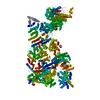
|
|---|---|
| 1 |
|
- Components
| #1: Protein | Mass: 527678.312 Da / Num. of mol.: 1 Source method: isolated from a genetically manipulated source Source: (gene. exp.)   Trichoplusia ni (cabbage looper) Trichoplusia ni (cabbage looper)References: UniProt: E9Q555, RING-type E3 ubiquitin transferase, Hydrolases; Acting on acid anhydrides; Acting on acid anhydrides to facilitate cellular and subcellular movement | ||
|---|---|---|---|
| #2: Chemical | ChemComp-ATP / | ||
| #3: Chemical | ChemComp-MG / | ||
| #4: Chemical | | Has ligand of interest | Y | |
-Experimental details
-Experiment
| Experiment | Method: ELECTRON MICROSCOPY |
|---|---|
| EM experiment | Aggregation state: PARTICLE / 3D reconstruction method: single particle reconstruction |
- Sample preparation
| Component | Name: full length RNF213 / Type: COMPLEX / Entity ID: #1 / Source: RECOMBINANT | ||||||||||||||||||||
|---|---|---|---|---|---|---|---|---|---|---|---|---|---|---|---|---|---|---|---|---|---|
| Molecular weight | Value: 0.58 MDa / Experimental value: YES | ||||||||||||||||||||
| Source (natural) | Organism:  | ||||||||||||||||||||
| Source (recombinant) | Organism:  Trichoplusia ni (cabbage looper) / Strain: BTI-Tn-5B1-4 / Plasmid: EMBacY Trichoplusia ni (cabbage looper) / Strain: BTI-Tn-5B1-4 / Plasmid: EMBacY | ||||||||||||||||||||
| Buffer solution | pH: 7.2 | ||||||||||||||||||||
| Buffer component |
| ||||||||||||||||||||
| Specimen | Conc.: 0.05 mg/ml / Embedding applied: NO / Shadowing applied: NO / Staining applied: NO / Vitrification applied: YES | ||||||||||||||||||||
| Specimen support | Details: SCD 005 Sputter Coater (BAL-TEC) grid sitting on a glass block during glow discharge Grid material: COPPER / Grid mesh size: 200 divisions/in. / Grid type: Quantifoil R2/2 | ||||||||||||||||||||
| Vitrification | Instrument: LEICA EM GP / Cryogen name: ETHANE / Chamber temperature: 277 K Details: sample incubated on the grid for 60 s prior to blotting. blotted for 2 seconds using using Whatman Filter Paper Grade 1 |
- Electron microscopy imaging
| Experimental equipment |  Model: Titan Krios / Image courtesy: FEI Company |
|---|---|
| Microscopy | Model: FEI TITAN KRIOS |
| Electron gun | Electron source:  FIELD EMISSION GUN / Accelerating voltage: 300 kV / Illumination mode: SPOT SCAN FIELD EMISSION GUN / Accelerating voltage: 300 kV / Illumination mode: SPOT SCAN |
| Electron lens | Mode: BRIGHT FIELD / Nominal magnification: 130000 X / Nominal defocus max: 3500 nm / Nominal defocus min: 1500 nm / Cs: 2.7 mm / C2 aperture diameter: 50 µm / Alignment procedure: COMA FREE |
| Specimen holder | Cryogen: NITROGEN / Specimen holder model: FEI TITAN KRIOS AUTOGRID HOLDER |
| Image recording | Average exposure time: 14 sec. / Electron dose: 58 e/Å2 / Detector mode: COUNTING / Film or detector model: GATAN K2 QUANTUM (4k x 4k) / Num. of grids imaged: 1 / Num. of real images: 4749 |
| EM imaging optics | Energyfilter name: GIF Bioquantum |
| Image scans | Width: 3838 / Height: 3710 / Movie frames/image: 40 / Used frames/image: 1-40 |
- Processing
| Software | Name: PHENIX / Version: 1.13_2998: / Classification: refinement | ||||||||||||||||||||||||||||||||||||||||||||||||
|---|---|---|---|---|---|---|---|---|---|---|---|---|---|---|---|---|---|---|---|---|---|---|---|---|---|---|---|---|---|---|---|---|---|---|---|---|---|---|---|---|---|---|---|---|---|---|---|---|---|
| EM software |
| ||||||||||||||||||||||||||||||||||||||||||||||||
| CTF correction | Type: PHASE FLIPPING AND AMPLITUDE CORRECTION | ||||||||||||||||||||||||||||||||||||||||||||||||
| Particle selection | Num. of particles selected: 375000 | ||||||||||||||||||||||||||||||||||||||||||||||||
| Symmetry | Point symmetry: C1 (asymmetric) | ||||||||||||||||||||||||||||||||||||||||||||||||
| 3D reconstruction | Resolution: 3.2 Å / Resolution method: FSC 0.143 CUT-OFF / Num. of particles: 374683 / Algorithm: FOURIER SPACE / Symmetry type: POINT | ||||||||||||||||||||||||||||||||||||||||||||||||
| Atomic model building | Protocol: AB INITIO MODEL / Space: REAL |
 Movie
Movie Controller


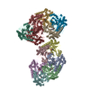
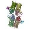
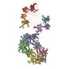
 PDBj
PDBj


