+ Open data
- Basic information
| Entry | Database: PDB / ID: 6t85 | ||||||||||||
|---|---|---|---|---|---|---|---|---|---|---|---|---|---|
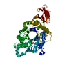
| Title | Urocanate reductase in complex with ADP | ||||||||||||
 Components Components | Urocanate reductase | ||||||||||||
 Keywords Keywords | OXIDOREDUCTASE / urocanate reductase / bacterial enzyme | ||||||||||||
| Function / homology |  Function and homology information Function and homology informationurocanate reductase / oxidoreductase activity, acting on the CH-CH group of donors / FMN binding / plasma membrane / cytoplasm Similarity search - Function | ||||||||||||
| Biological species |  Shewanella oneidensis (bacteria) Shewanella oneidensis (bacteria) | ||||||||||||
| Method |  X-RAY DIFFRACTION / X-RAY DIFFRACTION /  SYNCHROTRON / SYNCHROTRON /  MOLECULAR REPLACEMENT / MOLECULAR REPLACEMENT /  molecular replacement / Resolution: 1.1 Å molecular replacement / Resolution: 1.1 Å | ||||||||||||
 Authors Authors | Venskutonyte, R. / Lindkvist-Petersson, K. | ||||||||||||
| Funding support |  Sweden, 3items Sweden, 3items
| ||||||||||||
 Citation Citation |  Journal: Nat Commun / Year: 2021 Journal: Nat Commun / Year: 2021Title: Structural characterization of the microbial enzyme urocanate reductase mediating imidazole propionate production. Authors: Venskutonyte, R. / Koh, A. / Stenstrom, O. / Khan, M.T. / Lundqvist, A. / Akke, M. / Backhed, F. / Lindkvist-Petersson, K. | ||||||||||||
| History |
|
- Structure visualization
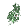
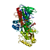
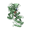
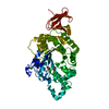
| Structure viewer | Molecule:  Molmil Molmil Jmol/JSmol Jmol/JSmol |
|---|
- Downloads & links
- Download
| PDBx/mmCIF format |  6t85.cif.gz 6t85.cif.gz | 316.9 KB | Display |  PDBx/mmCIF format PDBx/mmCIF format |
|---|---|---|---|---|
| PDB format |  pdb6t85.ent.gz pdb6t85.ent.gz | 259 KB | Display |  PDB format PDB format |
| PDBx/mmJSON format |  6t85.json.gz 6t85.json.gz | Tree view |  PDBx/mmJSON format PDBx/mmJSON format | |
| Others |  Other downloads Other downloads |
-Validation report
| Arichive directory |  https://data.pdbj.org/pub/pdb/validation_reports/t8/6t85 https://data.pdbj.org/pub/pdb/validation_reports/t8/6t85 ftp://data.pdbj.org/pub/pdb/validation_reports/t8/6t85 ftp://data.pdbj.org/pub/pdb/validation_reports/t8/6t85 | HTTPS FTP |
|---|
-Related structure data
| Related structure data |  6t86C  6t87C  6t88C 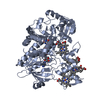 1d4cS S: Starting model for refinement C: citing same article ( |
|---|---|
| Similar structure data |
- Links
Links
- Assembly
| Deposited unit | 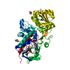
| ||||||||
|---|---|---|---|---|---|---|---|---|---|
| 1 |
| ||||||||
| Unit cell |
|
- Components
| #1: Protein | Mass: 49984.523 Da / Num. of mol.: 1 Source method: isolated from a genetically manipulated source Details: Truncated UrdA, construct comprising residues 130-582 and a C-terminal 6xHis tag. Source: (gene. exp.)  Shewanella oneidensis (strain MR-1) (bacteria) Shewanella oneidensis (strain MR-1) (bacteria)Strain: MR-1 / Gene: urdA, SO_4620 / Plasmid: pET-24a(+) / Production host:  | ||||||
|---|---|---|---|---|---|---|---|
| #2: Chemical | ChemComp-ADP / | ||||||
| #3: Chemical | ChemComp-CL / #4: Chemical | #5: Water | ChemComp-HOH / | Has ligand of interest | N | |
-Experimental details
-Experiment
| Experiment | Method:  X-RAY DIFFRACTION / Number of used crystals: 2 X-RAY DIFFRACTION / Number of used crystals: 2 |
|---|
- Sample preparation
| Crystal | Density Matthews: 2.53 Å3/Da / Density % sol: 51.43 % |
|---|---|
| Crystal grow | Temperature: 291 K / Method: vapor diffusion, hanging drop / pH: 7 / Details: 20 % PEG4000 0.1 M MgCl2 0.1 M HEPES pH 7.0. |
-Data collection
| Diffraction | Mean temperature: 100 K / Serial crystal experiment: N | ||||||||||||||||||||||||||||||
|---|---|---|---|---|---|---|---|---|---|---|---|---|---|---|---|---|---|---|---|---|---|---|---|---|---|---|---|---|---|---|---|
| Diffraction source | Source:  SYNCHROTRON / Site: SYNCHROTRON / Site:  PETRA III, EMBL c/o DESY PETRA III, EMBL c/o DESY  / Beamline: P13 (MX1) / Wavelength: 0.9762 Å / Beamline: P13 (MX1) / Wavelength: 0.9762 Å | ||||||||||||||||||||||||||||||
| Detector | Type: DECTRIS PILATUS3 6M / Detector: PIXEL / Date: Nov 30, 2018 | ||||||||||||||||||||||||||||||
| Radiation | Protocol: SINGLE WAVELENGTH / Monochromatic (M) / Laue (L): M / Scattering type: x-ray | ||||||||||||||||||||||||||||||
| Radiation wavelength | Wavelength: 0.9762 Å / Relative weight: 1 | ||||||||||||||||||||||||||||||
| Reflection | Resolution: 1.1→63.19 Å / Num. obs: 198264 / % possible obs: 98.6 % / Redundancy: 8.8 % / CC1/2: 0.999 / Rmerge(I) obs: 0.105 / Rpim(I) all: 0.036 / Rrim(I) all: 0.111 / Net I/σ(I): 9.5 / Num. measured all: 1753417 / Scaling rejects: 452 | ||||||||||||||||||||||||||||||
| Reflection shell | Diffraction-ID: 1
|
-Phasing
| Phasing | Method:  molecular replacement molecular replacement |
|---|
- Processing
| Software |
| ||||||||||||||||||||||||
|---|---|---|---|---|---|---|---|---|---|---|---|---|---|---|---|---|---|---|---|---|---|---|---|---|---|
| Refinement | Method to determine structure:  MOLECULAR REPLACEMENT MOLECULAR REPLACEMENTStarting model: 1D4C Resolution: 1.1→63.189 Å / SU ML: 0.1 / Cross valid method: THROUGHOUT / σ(F): 1.35 / Phase error: 12.1
| ||||||||||||||||||||||||
| Solvent computation | Shrinkage radii: 0.9 Å / VDW probe radii: 1.11 Å | ||||||||||||||||||||||||
| Displacement parameters | Biso max: 60.89 Å2 / Biso mean: 14.9925 Å2 / Biso min: 5.76 Å2 | ||||||||||||||||||||||||
| Refinement step | Cycle: final / Resolution: 1.1→63.189 Å
|
 Movie
Movie Controller





 PDBj
PDBj


