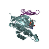
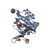
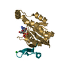
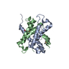
[English] 日本語
Yorodumi
- PDB-2qme: Crystal structure of human RAC3 in complex with CRIB domain of hu... -

+
Open data


ID or keywords:

Loading...

-
Basic information

Entry
Database: PDB / ID: 2qme
TitleCrystal structure of human RAC3 in complex with CRIB domain of human p21-activated kinase 1 (PAK1)
Components
  • CRIB domain of the Serine/threonine-protein kinase PAK 1
  • Ras-related C3 botulinum toxin substrate 3
KeywordsSIGNALING PROTEIN/TRANSFERASE / GTPase RAC3 / small GTP binding protein / p21 rac / ras-related C3 botulinum toxin substrate 3 / signaling protein / CRIB / kinase / Structural Genomics / Structural Genomics Consortium / SGC / Cell projection / GTP-binding / Lipoprotein / Membrane / Methylation / Nucleotide-binding / Prenylation / Allosteric enzyme / ATP-binding / Phosphorylation / Serine/threonine-protein kinase / Transferase / Apoptosis / Cell junction / SIGNALING PROTEIN-TRANSFERASE COMPLEX
Function / homology
Function and homology information


cerebral cortex GABAergic interneuron development / negative regulation of cell proliferation involved in contact inhibition / regulation of neuron maturation / protein localization to cytoplasmic stress granule / regulation of neutrophil migration / postsynaptic actin cytoskeleton organization / positive regulation of microtubule nucleation / engulfment of apoptotic cell / NADPH oxidase complex / respiratory burst ...cerebral cortex GABAergic interneuron development / negative regulation of cell proliferation involved in contact inhibition / regulation of neuron maturation / protein localization to cytoplasmic stress granule / regulation of neutrophil migration / postsynaptic actin cytoskeleton organization / positive regulation of microtubule nucleation / engulfment of apoptotic cell / NADPH oxidase complex / respiratory burst / cortical cytoskeleton organization / hepatocyte growth factor receptor signaling pathway / RHO GTPases Activate ROCKs / gamma-tubulin binding / cell projection assembly / synaptic transmission, GABAergic / motor neuron axon guidance / PCP/CE pathway / Activation of RAC1 / positive regulation of cell adhesion mediated by integrin / CD28 dependent Vav1 pathway / Ephrin signaling / positive regulation of intracellular estrogen receptor signaling pathway / RHOV GTPase cycle / establishment or maintenance of cell polarity / branching morphogenesis of an epithelial tube / regulation of axonogenesis / Fc-gamma receptor signaling pathway involved in phagocytosis / Rac protein signal transduction / RHOJ GTPase cycle / exocytosis / RHOQ GTPase cycle / filamentous actin / RHO GTPases activate PAKs / stimulatory C-type lectin receptor signaling pathway / regulation of MAPK cascade / CDC42 GTPase cycle / RHOU GTPase cycle / Sema3A PAK dependent Axon repulsion / neuromuscular process controlling balance / RHOH GTPase cycle / Generation of second messenger molecules / intercalated disc / Smooth Muscle Contraction / ephrin receptor signaling pathway / RAC2 GTPase cycle / RAC3 GTPase cycle / endomembrane system / positive regulation of substrate adhesion-dependent cell spreading / RHO GTPases activate PKNs / positive regulation of JUN kinase activity / homeostasis of number of cells within a tissue / positive regulation of stress fiber assembly / positive regulation of microtubule polymerization / ruffle / collagen binding / EPHB-mediated forward signaling / RAC1 GTPase cycle / CD209 (DC-SIGN) signaling / phosphorylation / cell chemotaxis / neuron projection morphogenesis / small monomeric GTPase / VEGFR2 mediated vascular permeability / G protein activity / Signal transduction by L1 / actin filament organization / cell periphery / actin filament / regulation of actin cytoskeleton organization / FCERI mediated MAPK activation / wound healing / MAPK6/MAPK4 signaling / G beta:gamma signalling through CDC42 / Regulation of actin dynamics for phagocytic cup formation / Wnt signaling pathway / ruffle membrane / Z disc / calcium-dependent protein binding / cell-cell junction / cell migration / positive regulation of peptidyl-serine phosphorylation / lamellipodium / chromosome / regulation of cell shape / growth cone / actin cytoskeleton organization / cytoplasmic vesicle / nuclear membrane / protein autophosphorylation / cytoskeleton / non-specific serine/threonine protein kinase / protein kinase activity / intracellular signal transduction / positive regulation of cell migration / neuron projection / chromatin remodeling / positive regulation of protein phosphorylation / axon / protein phosphorylation
Similarity search - Function
p21 activated kinase binding domain / : / CRIB domain superfamily / P21-Rho-binding domain / CRIB domain profile. / P21-Rho-binding domain / CRIB domain / Small GTPase Rho / small GTPase Rho family profile. / Rho (Ras homology) subfamily of Ras-like small GTPases ...p21 activated kinase binding domain / : / CRIB domain superfamily / P21-Rho-binding domain / CRIB domain profile. / P21-Rho-binding domain / CRIB domain / Small GTPase Rho / small GTPase Rho family profile. / Rho (Ras homology) subfamily of Ras-like small GTPases / Ras subfamily of RAS small GTPases / Small GTPase / Ras family / Rab subfamily of small GTPases / Small GTP-binding protein domain / P-loop containing nucleotide triphosphate hydrolases / Serine/threonine-protein kinase, active site / Serine/Threonine protein kinases active-site signature. / Protein kinase domain / Serine/Threonine protein kinases, catalytic domain / Protein kinase, ATP binding site / Protein kinases ATP-binding region signature. / Protein kinase domain profile. / Protein kinase domain / Protein kinase-like domain superfamily / Rossmann fold / P-loop containing nucleoside triphosphate hydrolase / 3-Layer(aba) Sandwich / Alpha Beta
Similarity search - Domain/homology
PHOSPHOMETHYLPHOSPHONIC ACID GUANYLATE ESTER / Ras-related C3 botulinum toxin substrate 3 / Serine/threonine-protein kinase PAK 1
Similarity search - Component
Biological speciesHomo sapiens (human)
MethodX-RAY DIFFRACTION / MOLECULAR REPLACEMENT / Resolution: 1.75 Å
AuthorsUgochukwu, E. / Yang, X. / Elkins, J.M. / Burgess-Brown, N. / Bunkoczi, G. / Sundstrom, M. / Arrowsmith, C.H. / Weigelt, J. / Edwards, A. / von Delft, F. ...Ugochukwu, E. / Yang, X. / Elkins, J.M. / Burgess-Brown, N. / Bunkoczi, G. / Sundstrom, M. / Arrowsmith, C.H. / Weigelt, J. / Edwards, A. / von Delft, F. / Knapp, S. / Doyle, D. / Structural Genomics Consortium (SGC)
CitationJournal: To be Published
Title: The crystal structure of the human RAC3 in complex with the CRIB domain of human p21-activated kinase 1 (PAK1).
Authors: Ugochukwu, E. / Yang, X. / Elkins, J.M. / Burgess-Brown, N. / Bunkoczi, G. / Knapp, S. / Doyle, D.
History
DepositionJul 16, 2007Deposition site: RCSB / Processing site: RCSB
Revision 1.0Aug 28, 2007Provider: repository / Type: Initial release
Revision 1.1Jul 13, 2011Group: Advisory / Version format compliance
Revision 1.2Aug 30, 2023Group: Data collection / Database references ...Data collection / Database references / Derived calculations / Refinement description
Category: chem_comp_atom / chem_comp_bond ...chem_comp_atom / chem_comp_bond / database_2 / pdbx_initial_refinement_model / pdbx_struct_conn_angle / struct_conn / struct_ref_seq_dif / struct_site
Item: _database_2.pdbx_DOI / _database_2.pdbx_database_accession ..._database_2.pdbx_DOI / _database_2.pdbx_database_accession / _pdbx_struct_conn_angle.ptnr1_auth_seq_id / _pdbx_struct_conn_angle.ptnr3_auth_seq_id / _pdbx_struct_conn_angle.value / _struct_conn.pdbx_dist_value / _struct_conn.ptnr2_auth_seq_id / _struct_ref_seq_dif.details / _struct_site.pdbx_auth_asym_id / _struct_site.pdbx_auth_comp_id / _struct_site.pdbx_auth_seq_id

-
Structure visualization

Structure viewerMolecule:
MolmilJmol/JSmol

Downloads & links

-
Assembly

Deposited unit
A: Ras-related C3 botulinum toxin substrate 3
I: CRIB domain of the Serine/threonine-protein kinase PAK 1
hetero molecules


Theoretical massNumber of molelcules
Total (without water)24,7286
Polymers23,9982
Non-polymers7304
Water4,288238
1


  • Idetical with deposited unit
  • defined by author&software
TypeNameSymmetry operationNumber
identity operation1_555x,y,z1
Buried area3240 Å2
MethodPISA
2
A: Ras-related C3 botulinum toxin substrate 3
I: CRIB domain of the Serine/threonine-protein kinase PAK 1
hetero molecules

A: Ras-related C3 botulinum toxin substrate 3
I: CRIB domain of the Serine/threonine-protein kinase PAK 1
hetero molecules


Theoretical massNumber of molelcules
Total (without water)49,45612
Polymers47,9974
Non-polymers1,4598
Water724
TypeNameSymmetry operationNumber
identity operation1_555x,y,z1
crystal symmetry operation2_556-x,y,-z+11
Buried area8640 Å2
MethodPISA
Unit cell
Length a, b, c (Å)84.226, 53.480, 47.682
Angle α, β, γ (deg.)90.00, 93.45, 90.00
Int Tables number5
Space group name H-MC121
Components on special symmetry positions
IDModelComponents
11A-305-

HOH

21A-511-

HOH

-
Components

-
Protein / Protein/peptide , 2 types, 2 molecules AI

#1: Protein Ras-related C3 botulinum toxin substrate 3 / p21-Rac3


Mass: 19934.928 Da / Num. of mol.: 1
Source method: isolated from a genetically manipulated source
Source: (gene. exp.) Homo sapiens (human) / Gene: RAC3 / Plasmid: pNIC28-Bsa4 / Production host: Escherichia coli (E. coli) / Strain (production host): BL21(DE3)-R3 / References: UniProt: P60763
#2: Protein/peptide CRIB domain of the Serine/threonine-protein kinase PAK 1 / p21-activated kinase 1 / PAK-1 / p65-PAK / Alpha-PAK


Mass: 4063.479 Da / Num. of mol.: 1 / Fragment: CRIB domain / Source method: obtained synthetically
Details: Synthetic peptide with the sequence of the CRIB domain of human PAK1 protein
References: UniProt: Q13153, non-specific serine/threonine protein kinase

-
Non-polymers , 4 types, 242 molecules

#3: Chemical ChemComp-MG / MAGNESIUM ION


Mass: 24.305 Da / Num. of mol.: 1 / Source method: obtained synthetically / Formula: Mg
#4: Chemical ChemComp-GCP / PHOSPHOMETHYLPHOSPHONIC ACID GUANYLATE ESTER


Mass: 521.208 Da / Num. of mol.: 1 / Source method: obtained synthetically / Formula: C11H18N5O13P3 / Comment: GMP-PCP, energy-carrying molecule analogue*YM
#5: Chemical ChemComp-GOL / GLYCEROL / GLYCERIN / PROPANE-1,2,3-TRIOL


Mass: 92.094 Da / Num. of mol.: 2 / Source method: obtained synthetically / Formula: C3H8O3
#6: Water ChemComp-HOH / water


Mass: 18.015 Da / Num. of mol.: 238 / Source method: isolated from a natural source / Formula: H2O

-
Experimental details

-
Experiment

ExperimentMethod: X-RAY DIFFRACTION / Number of used crystals: 1

-
Sample preparation

CrystalDensity Matthews: 2.23 Å3/Da / Density % sol: 44.93 %
Crystal growTemperature: 293 K / Method: vapor diffusion, sitting drop / pH: 8.5
Details: 0.1 M Tris-HCl pH 8.5, 2 M (NH4)2S04, VAPOR DIFFUSION, SITTING DROP, temperature 293K

-
Data collection

DiffractionMean temperature: 100 K
Diffraction sourceSource: ROTATING ANODE / Type: RIGAKU FR-E+ SUPERBRIGHT / Wavelength: 1.5418 Å
DetectorType: RIGAKU RAXIS HTC / Detector: IMAGE PLATE / Date: Jan 31, 2007
RadiationProtocol: SINGLE WAVELENGTH / Monochromatic (M) / Laue (L): M / Scattering type: x-ray
Radiation wavelengthWavelength: 1.5418 Å / Relative weight: 1
ReflectionResolution: 1.75→47.62 Å / Num. all: 21448 / Num. obs: 21448 / % possible obs: 99.9 % / Observed criterion σ(F): 0 / Observed criterion σ(I): 0 / Redundancy: 7.2 % / Biso Wilson estimate: 20 Å2 / Rmerge(I) obs: 0.053 / Rsym value: 0.053 / Net I/σ(I): 27.1
Reflection shellResolution: 1.75→1.84 Å / Redundancy: 7 % / Rmerge(I) obs: 0.343 / Mean I/σ(I) obs: 4 / Rsym value: 0.343 / % possible all: 99.6

-
Processing

Software
NameVersionClassification
REFMAC5.3.0037refinement
CrystalCleardata collection
MOSFLMdata reduction
SCALAdata scaling
PHASERphasing
RefinementMethod to determine structure: MOLECULAR REPLACEMENT
Starting model: PDB entry 2G0N
Resolution: 1.75→47.62 Å / Cor.coef. Fo:Fc: 0.969 / Cor.coef. Fo:Fc free: 0.95 / SU B: 3.899 / SU ML: 0.069 / TLS residual ADP flag: LIKELY RESIDUAL / Cross valid method: THROUGHOUT / σ(F): 0 / ESU R: 0.101 / ESU R Free: 0.103 / Stereochemistry target values: MAXIMUM LIKELIHOOD / Details: HYDROGENS HAVE BEEN ADDED IN THE RIDING POSITIONS
RfactorNum. reflection% reflectionSelection details
Rfree0.19177 1100 5.1 %RANDOM
Rwork0.14893 ---
all0.15114 20345 --
obs0.15114 20345 99.88 %-
Solvent computationIon probe radii: 0.8 Å / Shrinkage radii: 0.8 Å / VDW probe radii: 1.2 Å / Solvent model: BABINET MODEL WITH MASK
Displacement parametersBiso mean: 22.09 Å2
Baniso -1Baniso -2Baniso -3
1--0.15 Å20 Å20.02 Å2
2--0.59 Å20 Å2
3----0.43 Å2
Refinement stepCycle: LAST / Resolution: 1.75→47.62 Å
ProteinNucleic acidLigandSolventTotal
Num. atoms1560 0 45 238 1843
Refine LS restraints
Refine-IDTypeDev idealDev ideal targetNumber
X-RAY DIFFRACTIONr_bond_refined_d0.0130.0221663
X-RAY DIFFRACTIONr_bond_other_d0.0020.021095
X-RAY DIFFRACTIONr_angle_refined_deg1.471.9962273
X-RAY DIFFRACTIONr_angle_other_deg0.88432681
X-RAY DIFFRACTIONr_dihedral_angle_1_deg5.9175202
X-RAY DIFFRACTIONr_dihedral_angle_2_deg38.21524.05869
X-RAY DIFFRACTIONr_dihedral_angle_3_deg11.61215258
X-RAY DIFFRACTIONr_dihedral_angle_4_deg11.228158
X-RAY DIFFRACTIONr_chiral_restr0.0880.2257
X-RAY DIFFRACTIONr_gen_planes_refined0.0060.021808
X-RAY DIFFRACTIONr_gen_planes_other0.0010.02329
X-RAY DIFFRACTIONr_nbd_refined0.2020.2336
X-RAY DIFFRACTIONr_nbd_other0.2020.21182
X-RAY DIFFRACTIONr_nbtor_refined0.1810.2816
X-RAY DIFFRACTIONr_nbtor_other0.0850.2806
X-RAY DIFFRACTIONr_xyhbond_nbd_refined0.1680.2168
X-RAY DIFFRACTIONr_metal_ion_refined0.0210.21
X-RAY DIFFRACTIONr_symmetry_vdw_refined0.1710.215
X-RAY DIFFRACTIONr_symmetry_vdw_other0.2580.247
X-RAY DIFFRACTIONr_symmetry_hbond_refined0.1350.230
X-RAY DIFFRACTIONr_mcbond_it3.51751051
X-RAY DIFFRACTIONr_mcbond_other1.2865405
X-RAY DIFFRACTIONr_mcangle_it4.29571644
X-RAY DIFFRACTIONr_scbond_it6.1759728
X-RAY DIFFRACTIONr_scangle_it7.43511628
LS refinement shellResolution: 1.75→1.795 Å / Total num. of bins used: 20
RfactorNum. reflection% reflection
Rfree0.321 86 -
Rwork0.194 1481 -
obs--99.18 %
Refinement TLS params.Method: refined / Origin x: -12.6483 Å / Origin y: 12.2223 Å / Origin z: 13.6153 Å
111213212223313233
T-0.0315 Å2-0.0033 Å20.0029 Å2--0.0232 Å2-0.0003 Å2---0.0216 Å2
L0.4667 °20.0262 °20.1486 °2-0.5499 °20.3052 °2--0.9467 °2
S-0.0004 Å °0.0249 Å °-0.0318 Å °0.0024 Å °0.0498 Å °-0.0251 Å °-0.0216 Å °0.0492 Å °-0.0494 Å °
Refinement TLS group
IDRefine-IDRefine TLS-IDAuth asym-IDLabel asym-IDAuth seq-IDLabel seq-ID
1X-RAY DIFFRACTION1AA1 - 1782 - 179
2X-RAY DIFFRACTION1IB10 - 331 - 24

+
About Yorodumi

-
News

-
Feb 9, 2022. New format data for meta-information of EMDB entries

New format data for meta-information of EMDB entries

  • Version 3 of the EMDB header file is now the official format.
  • The previous official version 1.9 will be removed from the archive.

Related info.:EMDB header

External links:wwPDB to switch to version 3 of the EMDB data model

-
Aug 12, 2020. Covid-19 info

Covid-19 info

URL: https://pdbjlc1.pdbj.org/emnavi/covid19.php

New page: Covid-19 featured information page in EM Navigator.

Related info.:Covid-19 info / Mar 5, 2020. Novel coronavirus structure data

+
Mar 5, 2020. Novel coronavirus structure data

Novel coronavirus structure data

Related info.:Yorodumi Speices / Aug 12, 2020. Covid-19 info

External links:COVID-19 featured content - PDBj / Molecule of the Month (242):Coronavirus Proteases

+
Jan 31, 2019. EMDB accession codes are about to change! (news from PDBe EMDB page)

EMDB accession codes are about to change! (news from PDBe EMDB page)

  • The allocation of 4 digits for EMDB accession codes will soon come to an end. Whilst these codes will remain in use, new EMDB accession codes will include an additional digit and will expand incrementally as the available range of codes is exhausted. The current 4-digit format prefixed with “EMD-” (i.e. EMD-XXXX) will advance to a 5-digit format (i.e. EMD-XXXXX), and so on. It is currently estimated that the 4-digit codes will be depleted around Spring 2019, at which point the 5-digit format will come into force.
  • The EM Navigator/Yorodumi systems omit the EMD- prefix.

Related info.:Q: What is EMD? / ID/Accession-code notation in Yorodumi/EM Navigator

External links:EMDB Accession Codes are Changing Soon! / Contact to PDBj

+
Jul 12, 2017. Major update of PDB

Major update of PDB

  • wwPDB released updated PDB data conforming to the new PDBx/mmCIF dictionary.
  • This is a major update changing the version number from 4 to 5, and with Remediation, in which all the entries are updated.
  • In this update, many items about electron microscopy experimental information are reorganized (e.g. em_software).
  • Now, EM Navigator and Yorodumi are based on the updated data.

External links:wwPDB Remediation / Enriched Model Files Conforming to OneDep Data Standards Now Available in the PDB FTP Archive

-
Yorodumi

Thousand views of thousand structures

  • Yorodumi is a browser for structure data from EMDB, PDB, SASBDB, etc.
  • This page is also the successor to EM Navigator detail page, and also detail information page/front-end page for Omokage search.
  • The word "yorodu" (or yorozu) is an old Japanese word meaning "ten thousand". "mi" (miru) is to see.

Related info.:EMDB / PDB / SASBDB / Comparison of 3 databanks / Yorodumi Search / Aug 31, 2016. New EM Navigator & Yorodumi / Yorodumi Papers / Jmol/JSmol / Function and homology information / Changes in new EM Navigator and Yorodumi

Read more