 Movie
Movie Controller

[English] 日本語
 Yorodumi
- PDB-6t77: Crystal structure of Klebsiella pneumoniae FabG(NADPH-dependent) ... -
+ Open data

- Basic information
| Entry | Database: PDB / ID: 6t77 | ||||||
|---|---|---|---|---|---|---|---|
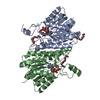
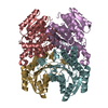
| Title | Crystal structure of Klebsiella pneumoniae FabG(NADPH-dependent) NADP-complex at 1.75 A resolution | ||||||
 Components Components | 3-oxoacyl-ACP reductase | ||||||
 Keywords Keywords | BIOSYNTHETIC PROTEIN / Fatty acid biosynthesis / FabG / (3-oxoacyl-(Acyl-carrier-protein) reductase) / NADPH / complex / FAS-II | ||||||
| Function / homology |  Function and homology information Function and homology information3-oxoacyl-[acyl-carrier-protein] reductase / 3-oxoacyl-[acyl-carrier-protein] reductase (NADPH) activity / NAD binding / fatty acid biosynthetic process Similarity search - Function | ||||||
| Biological species |  Klebsiella pneumoniae (bacteria) Klebsiella pneumoniae (bacteria) | ||||||
| Method |  X-RAY DIFFRACTION / SYNCHROTRON / MOLECULAR REPLACEMENT / Resolution: 1.75 Å X-RAY DIFFRACTION / SYNCHROTRON / MOLECULAR REPLACEMENT / Resolution: 1.75 Å | ||||||
 Authors Authors | Vella, P. / Schnell, R. / Lindqvist, Y. / Schneider, G. | ||||||
| Funding support |  Sweden, 1items Sweden, 1items
| ||||||
 Citation Citation | Journal: Bioorg.Med.Chem. / Year: 2021 Title: A FabG inhibitor targeting an allosteric binding site inhibits several orthologs from Gram-negative ESKAPE pathogens. Authors: Vella, P. / Rudraraju, R.S. / Lundback, T. / Axelsson, H. / Almqvist, H. / Vallin, M. / Schneider, G. / Schnell, R. | ||||||
| History |
|
- Structure visualization
| Structure viewer | Molecule: MolmilJmol/JSmol |
|---|
- Downloads & links
-Download
| PDBx/mmCIF format | 6t77.cif.gz | 200.5 KB | Display | PDBx/mmCIF format |
|---|---|---|---|---|
| PDB format | pdb6t77.ent.gz | 160.6 KB | Display | PDB format |
| PDBx/mmJSON format | 6t77.json.gz | Tree view | PDBx/mmJSON format | |
| Others |  Other downloads Other downloads |
-Validation report
| Arichive directory | https://data.pdbj.org/pub/pdb/validation_reports/t7/6t77ftp://data.pdbj.org/pub/pdb/validation_reports/t7/6t77 | HTTPS FTP |
|---|
-Related structure data
| Related structure data |  6t5xC  6t60C  6t62C  6t65C  6t6nC  6t6pC  6t7mC  1q7bS S: Starting model for refinement C: citing same article ( |
|---|---|
| Similar structure data |


-Links
 PDBj
PDBj


- Assembly
| Deposited unit | 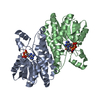
| ||||||||||||||||||
|---|---|---|---|---|---|---|---|---|---|---|---|---|---|---|---|---|---|---|---|
| 1 | 
| ||||||||||||||||||
| Unit cell |
| ||||||||||||||||||
| Noncrystallographic symmetry (NCS) | NCS domain:
NCS domain segments: Component-ID: _ / Ens-ID: 1 / Beg auth comp-ID: MET / Beg label comp-ID: MET / End auth comp-ID: VAL / End label comp-ID: VAL / Refine code: _ / Auth seq-ID: 1 - 244 / Label seq-ID: 2 - 245
|
-Components
| #1: Protein | Mass: 25623.238 Da / Num. of mol.: 2 Source method: isolated from a genetically manipulated source Details: The protein was expressed in E.coli with N-terminal His6-tag (removable by TEV protease cleavage). The tag removal results in a protein with one extra Serine residue at the N-terminus. Source: (gene. exp.) Klebsiella pneumoniae (bacteria)Gene: fabG, fabG_1, fabG_10, fabG_11, fabG_13, fabG_17, fabG_2, fabG_20, fabG_29, fabG_3, fabG_5, fabG_6, fabG_7, fabG_9, B4U21_08660, B4U25_10170, B4U30_10945, BANRA_00187, BANRA_00827, BL124_ ...Gene: fabG, fabG_1, fabG_10, fabG_11, fabG_13, fabG_17, fabG_2, fabG_20, fabG_29, fabG_3, fabG_5, fabG_6, fabG_7, fabG_9, B4U21_08660, B4U25_10170, B4U30_10945, BANRA_00187, BANRA_00827, BL124_00014385, BN49_2178, BU230_37105, BVX91_09955, C1459_07275, C3483_16280, C3F39_04320, C4Y50_026525, C7V41_16750, C8P71_1985, CSC88_11955, CWN54_18865, DD581_15755, DD583_12070, DM059_09455, DN589_06290, DXF97_11560, E0760_18120, EAO17_24465, EXT45_09615, FAQ72_04165, FAQ97_22680, FAS39_22705, NCTC11679_03520, NCTC13465_02289, NCTC13635_06738, NCTC1936_03302, NCTC204_06212, NCTC5052_01093, NCTC8849_02353, NCTC9128_07518, NCTC9178_04075, NCTC9617_01456, NCTC9637_04333, NCTC9645_00460, PMK1_03430, SAMEA104305404_12205, SAMEA104567806_03967, SAMEA104567857_00497, SAMEA104567903_00037, SAMEA23986918_09908, SAMEA3531848_01000, SAMEA4364603_00036, SAMEA4394730_07307, SAMEA4873640_04565, SK89_00756, SM87_01800 Plasmid: pNIC28Bsa4 Details (production host): N-terminal His6-tag (removable by TEV protease cleavage) Production host: References: UniProt: W9B6I8, 3-oxoacyl-[acyl-carrier-protein] reductase #2: Chemical |   Mass: 743.405 Da / Num. of mol.: 2 / Source method: obtained synthetically / Formula: C21H28N7O17P3 Mass: 743.405 Da / Num. of mol.: 2 / Source method: obtained synthetically / Formula: C21H28N7O17P3Details: In chain A it is partialy disordered, and only the Pho-Adenine nucleotide segment is resolved in the electron density map. Feature type: SUBJECT OF INVESTIGATION #3: Water | ChemComp-HOH / |  Mass: 18.015 Da / Num. of mol.: 217 / Source method: isolated from a natural source / Formula: H2O Mass: 18.015 Da / Num. of mol.: 217 / Source method: isolated from a natural source / Formula: H2OHas ligand of interest | Y | |
|---|
-Experimental details
-Experiment
| Experiment | Method: X-RAY DIFFRACTION / Number of used crystals: 1 |
|---|
- Sample preparation
| Crystal | Density Matthews: 2.39 Å3/Da / Density % sol: 48.44 % |
|---|---|
| Crystal grow | Temperature: 297 K / Method: vapor diffusion, sitting drop / pH: 4.2 Details: 9.5 mg/ml protein solution mixed with NADP+ at 11.5 mM concentration Well solution: 0.1 M phosphate-citrate 4.2 pH 40 %v/v PEG 300 |
-Data collection
| Diffraction | Mean temperature: 100 K / Serial crystal experiment: N |
|---|---|
| Diffraction source | Source: SYNCHROTRON / Site: ESRF  / Beamline: ID23-1 / Wavelength: 0.96862 Å / Beamline: ID23-1 / Wavelength: 0.96862 Å |
| Detector | Type: DECTRIS PILATUS3 S 6M / Detector: PIXEL / Date: Jun 17, 2017 / Details: Toroidal mirrors |
| Radiation | Monochromator: Si (111) / Protocol: SINGLE WAVELENGTH / Monochromatic (M) / Laue (L): M / Scattering type: x-ray |
| Radiation wavelength | Wavelength: 0.96862 Å / Relative weight: 1 |
| Reflection | Resolution: 1.75→59.88 Å / Num. obs: 47966 / % possible obs: 96.8 % / Redundancy: 3.8 % / Biso Wilson estimate: 27.9 Å2 / CC1/2: 0.998 / Rmerge(I) obs: 0.034 / Rpim(I) all: 0.026 / Net I/σ(I): 15 |
| Reflection shell | Resolution: 1.75→1.78 Å / Redundancy: 3.8 % / Rmerge(I) obs: 0.38 / Mean I/σ(I) obs: 2 / Num. unique obs: 2647 / CC1/2: 0.861 / Rpim(I) all: 0.297 / % possible all: 99.3 |
- Processing
| Software |
| |||||||||||||||||||||||||||||||||||||||||||||||||||||||||||||||||||||||||||
|---|---|---|---|---|---|---|---|---|---|---|---|---|---|---|---|---|---|---|---|---|---|---|---|---|---|---|---|---|---|---|---|---|---|---|---|---|---|---|---|---|---|---|---|---|---|---|---|---|---|---|---|---|---|---|---|---|---|---|---|---|---|---|---|---|---|---|---|---|---|---|---|---|---|---|---|---|
| Refinement | Method to determine structure: MOLECULAR REPLACEMENT Starting model: 1Q7B Resolution: 1.75→59.88 Å / Cor.coef. Fo:Fc: 0.976 / Cor.coef. Fo:Fc free: 0.963 / Cross valid method: THROUGHOUT / σ(F): 0 / ESU R: 0.103 / ESU R Free: 0.103 Details: HYDROGENS HAVE BEEN ADDED IN THE RIDING POSITIONS U VALUES : WITH TLS ADDED
| |||||||||||||||||||||||||||||||||||||||||||||||||||||||||||||||||||||||||||
| Solvent computation | Ion probe radii: 0.8 Å / Shrinkage radii: 0.8 Å / VDW probe radii: 1.2 Å | |||||||||||||||||||||||||||||||||||||||||||||||||||||||||||||||||||||||||||
| Displacement parameters | Biso max: 108.71 Å2 / Biso mean: 38.978 Å2 / Biso min: 21.55 Å2
| |||||||||||||||||||||||||||||||||||||||||||||||||||||||||||||||||||||||||||
| Refinement step | Cycle: final / Resolution: 1.75→59.88 Å
| |||||||||||||||||||||||||||||||||||||||||||||||||||||||||||||||||||||||||||
| Refine LS restraints |
| |||||||||||||||||||||||||||||||||||||||||||||||||||||||||||||||||||||||||||
| Refine LS restraints NCS | Ens-ID: 1 / Number: 14464 / Refine-ID: X-RAY DIFFRACTION / Type: interatomic distance / Rms dev position: 0.07 Å / Weight position: 0.05
| |||||||||||||||||||||||||||||||||||||||||||||||||||||||||||||||||||||||||||
| LS refinement shell | Resolution: 1.75→1.795 Å / Rfactor Rfree error: 0 / Total num. of bins used: 20
| |||||||||||||||||||||||||||||||||||||||||||||||||||||||||||||||||||||||||||
| Refinement TLS params. | Method: refined / Refine-ID: X-RAY DIFFRACTION
| |||||||||||||||||||||||||||||||||||||||||||||||||||||||||||||||||||||||||||
| Refinement TLS group |
|
