 Movie
Movie Controller

[English] 日本語
 Yorodumi
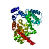
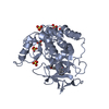
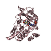
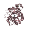
- PDB-6szo: The glucuronoyl esterase OtCE15A S267A variant from Opitutus terr... -
+ Open data

- Basic information
| Entry | Database: PDB / ID: 6szo | |||||||||
|---|---|---|---|---|---|---|---|---|---|---|
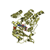
| Title | The glucuronoyl esterase OtCE15A S267A variant from Opitutus terrae in complex with D-galacturonate | |||||||||
 Components Components | glucuronoyl esterase OtCE15A | |||||||||
 Keywords Keywords | HYDROLASE / Esterase / Complex / Biomass | |||||||||
| Function / homology | : / Glucuronyl esterase, fungi / carboxylic ester hydrolase activity / Alpha/Beta hydrolase fold / metal ion binding / beta-D-galactopyranuronic acid / DI(HYDROXYETHYL)ETHER / TRIETHYLENE GLYCOL / Putative acetyl xylan esterase Function and homology information Function and homology information | |||||||||
| Biological species |  Opitutus terrae PB90-1 (bacteria) Opitutus terrae PB90-1 (bacteria) | |||||||||
| Method |  X-RAY DIFFRACTION / SYNCHROTRON / MOLECULAR REPLACEMENT / Resolution: 2.2 Å X-RAY DIFFRACTION / SYNCHROTRON / MOLECULAR REPLACEMENT / Resolution: 2.2 Å | |||||||||
 Authors Authors | Mazurkewich, S. / Navarro Poulsen, J.C. / Larsbrink, J. / Lo Leggio, L. | |||||||||
| Funding support |  Sweden, Sweden,  Denmark, 2items Denmark, 2items
| |||||||||
 Citation Citation | Journal: J.Biol.Chem. / Year: 2019 Title: Structural and biochemical studies of the glucuronoyl esteraseOtCE15A illuminate its interaction with lignocellulosic components. Authors: Mazurkewich, S. / Poulsen, J.N. / Lo Leggio, L. / Larsbrink, J. | |||||||||
| History |
|
- Structure visualization
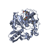
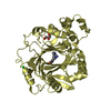
| Structure viewer | Molecule: MolmilJmol/JSmol |
|---|
- Downloads & links
-Download
| PDBx/mmCIF format | 6szo.cif.gz | 159.6 KB | Display | PDBx/mmCIF format |
|---|---|---|---|---|
| PDB format | pdb6szo.ent.gz | 124.1 KB | Display | PDB format |
| PDBx/mmJSON format | 6szo.json.gz | Tree view | PDBx/mmJSON format | |
| Others |  Other downloads Other downloads |
-Validation report
| Summary document | 6szo_validation.pdf.gz | 847.4 KB | Display | wwPDB validaton report |
|---|---|---|---|---|
| Full document | 6szo_full_validation.pdf.gz | 852.7 KB | Display | |
| Data in XML | 6szo_validation.xml.gz | 17.9 KB | Display | |
| Data in CIF | 6szo_validation.cif.gz | 25 KB | Display | |
| Arichive directory | https://data.pdbj.org/pub/pdb/validation_reports/sz/6szoftp://data.pdbj.org/pub/pdb/validation_reports/sz/6szo | HTTPS FTP |
-Related structure data
| Related structure data |  6syrC  6syuC  6syvC  6sz0C 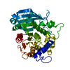 6sz4C  6t0eC  6t0iC  6gs0S S: Starting model for refinement C: citing same article ( |
|---|---|
| Similar structure data |
-Links
 PDBj
PDBj- Assembly
| Deposited unit | 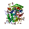
| ||||||||||||
|---|---|---|---|---|---|---|---|---|---|---|---|---|---|
| 1 |
| ||||||||||||
| Unit cell |
|
-Components
-Protein / Sugars , 2 types, 2 molecules A
| #1: Protein | Mass: 46132.547 Da / Num. of mol.: 1 / Mutation: S267A Source method: isolated from a genetically manipulated source Source: (gene. exp.) Opitutus terrae PB90-1 (bacteria) / Gene: Oter_0116 / Production host: |
|---|---|
| #2: Sugar | ChemComp-GTR /  Type: D-saccharide, beta linking / Mass: 194.139 Da / Num. of mol.: 1 Type: D-saccharide, beta linking / Mass: 194.139 Da / Num. of mol.: 1Source method: isolated from a genetically manipulated source Formula: C6H10O7 / Feature type: SUBJECT OF INVESTIGATION |
-Non-polymers , 6 types, 159 molecules 


| #3: Chemical | ChemComp-DMS /  Mass: 78.133 Da / Num. of mol.: 4 / Source method: obtained synthetically / Formula: C2H6OS / Comment: DMSO, precipitant*YM Mass: 78.133 Da / Num. of mol.: 4 / Source method: obtained synthetically / Formula: C2H6OS / Comment: DMSO, precipitant*YM#4: Chemical | ChemComp-MG / |  Mass: 24.305 Da / Num. of mol.: 1 / Source method: obtained synthetically / Formula: Mg Mass: 24.305 Da / Num. of mol.: 1 / Source method: obtained synthetically / Formula: Mg#5: Chemical | ChemComp-EDO /  Mass: 62.068 Da / Num. of mol.: 24 / Source method: obtained synthetically / Formula: C2H6O2 Mass: 62.068 Da / Num. of mol.: 24 / Source method: obtained synthetically / Formula: C2H6O2#6: Chemical | ChemComp-PEG /  Mass: 106.120 Da / Num. of mol.: 4 / Source method: obtained synthetically / Formula: C4H10O3 Mass: 106.120 Da / Num. of mol.: 4 / Source method: obtained synthetically / Formula: C4H10O3#7: Chemical | ChemComp-PGE /  Mass: 150.173 Da / Num. of mol.: 4 / Source method: obtained synthetically / Formula: C6H14O4 Mass: 150.173 Da / Num. of mol.: 4 / Source method: obtained synthetically / Formula: C6H14O4#8: Water | ChemComp-HOH / | Mass: 18.015 Da / Num. of mol.: 122 / Source method: isolated from a natural source / Formula: H2O |
|---|
-Details
| Has ligand of interest | Y |
|---|
-Experimental details
-Experiment
| Experiment | Method: X-RAY DIFFRACTION / Number of used crystals: 1 |
|---|
- Sample preparation
| Crystal | Density Matthews: 1.89 Å3/Da / Density % sol: 34.92 % |
|---|---|
| Crystal grow | Temperature: 298 K / Method: vapor diffusion, sitting drop / pH: 7.5 Details: Enzyme mixed 50/50 with reservoir solution containing Morpheus screen solution E8: 0.12 M Ethylene glycols (0.3M Diethylene glycol; 0.3M Triethylene glycol; 0.3M Tetraethylene glycol; 0.3M ...Details: Enzyme mixed 50/50 with reservoir solution containing Morpheus screen solution E8: 0.12 M Ethylene glycols (0.3M Diethylene glycol; 0.3M Triethylene glycol; 0.3M Tetraethylene glycol; 0.3M Pentaethylene glycol), 0.1 M Buffer System 2 pH 7.5 (Sodium HEPES; MOPS), and 50 % v/v Precipitant Mix 4 (25% v/v MPD; 25% PEG 1000; 25% w/v PEG 3350) |
-Data collection
| Diffraction | Mean temperature: 100 K / Serial crystal experiment: N |
|---|---|
| Diffraction source | Source: SYNCHROTRON / Site: PETRA III, DESY  / Beamline: P11 / Wavelength: 0.9891 Å / Beamline: P11 / Wavelength: 0.9891 Å |
| Detector | Type: DECTRIS PILATUS 6M / Detector: PIXEL / Date: Sep 18, 2018 |
| Radiation | Protocol: SINGLE WAVELENGTH / Monochromatic (M) / Laue (L): M / Scattering type: x-ray |
| Radiation wavelength | Wavelength: 0.9891 Å / Relative weight: 1 |
| Reflection | Resolution: 2.198→44.74 Å / Num. obs: 16711 / % possible obs: 97.04 % / Redundancy: 2.7 % / Biso Wilson estimate: 30.4 Å2 / CC1/2: 0.996 / Rmerge(I) obs: 0.08127 / Rpim(I) all: 0.05835 / Rrim(I) all: 0.1006 / Net I/σ(I): 8.66 |
| Reflection shell | Resolution: 2.198→2.277 Å / Redundancy: 2 % / Rmerge(I) obs: 0.4798 / Mean I/σ(I) obs: 2 / Num. unique obs: 1620 / CC1/2: 0.747 / Rpim(I) all: 0.3448 / Rrim(I) all: 0.594 / % possible all: 94.62 |
- Processing
| Software |
| ||||||||||||||||||||||||||||||||||||||||||||||||||||||||||||||||||||||
|---|---|---|---|---|---|---|---|---|---|---|---|---|---|---|---|---|---|---|---|---|---|---|---|---|---|---|---|---|---|---|---|---|---|---|---|---|---|---|---|---|---|---|---|---|---|---|---|---|---|---|---|---|---|---|---|---|---|---|---|---|---|---|---|---|---|---|---|---|---|---|---|
| Refinement | Method to determine structure: MOLECULAR REPLACEMENT Starting model: 6gs0 Resolution: 2.2→44.74 Å / SU ML: 0.2418 / Cross valid method: FREE R-VALUE / σ(F): 1.98 / Phase error: 23.5455
| ||||||||||||||||||||||||||||||||||||||||||||||||||||||||||||||||||||||
| Solvent computation | Shrinkage radii: 0.9 Å / VDW probe radii: 1.11 Å | ||||||||||||||||||||||||||||||||||||||||||||||||||||||||||||||||||||||
| Displacement parameters | Biso mean: 35.1 Å2 | ||||||||||||||||||||||||||||||||||||||||||||||||||||||||||||||||||||||
| Refinement step | Cycle: LAST / Resolution: 2.2→44.74 Å
| ||||||||||||||||||||||||||||||||||||||||||||||||||||||||||||||||||||||
| Refine LS restraints |
| ||||||||||||||||||||||||||||||||||||||||||||||||||||||||||||||||||||||
| LS refinement shell |
|
