 Movie
Movie Controller


+ Open data

- Basic information
| Entry | Database: PDB / ID: 6sym | ||||||
|---|---|---|---|---|---|---|---|
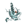
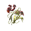
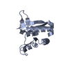
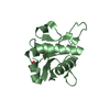
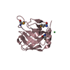
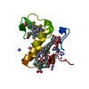
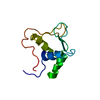
| Title | Crystal structure of Escherichia coli MsrB (reduced form) | ||||||
 Components Components | Peptide methionine sulfoxide reductase MsrB | ||||||
 Keywords Keywords | OXIDOREDUCTASE / Reductase / Zinc-binding / R-Methionine sulfoxide reductase / Oxidative stress | ||||||
| Function / homology |  Function and homology information Function and homology informationL-methionine (R)-S-oxide reductase activity / peptide-methionine (R)-S-oxide reductase / peptide-methionine (R)-S-oxide reductase activity / protein repair / response to cadmium ion / response to oxidative stress / iron ion binding / zinc ion binding / cytoplasm / cytosol Similarity search - Function | ||||||
| Biological species |  | ||||||
| Method |  X-RAY DIFFRACTION / SYNCHROTRON / MOLECULAR REPLACEMENT / molecular replacement / Resolution: 1.6302 Å X-RAY DIFFRACTION / SYNCHROTRON / MOLECULAR REPLACEMENT / molecular replacement / Resolution: 1.6302 Å | ||||||
 Authors Authors | Napolitano, S. / Zyla, D. / Glockshuber, R. | ||||||
 Citation Citation | Journal: To Be Published Title: Structure of Peptide methionine sulfoxide reductase MsrB at 1.63 Angstrom resolution Authors: Napolitano, S. / Zyla, D. / Glockshuber, R. | ||||||
| History |
|
- Structure visualization
| Structure viewer | Molecule: MolmilJmol/JSmol |
|---|
- Downloads & links
-Download
| PDBx/mmCIF format | 6sym.cif.gz | 164.4 KB | Display | PDBx/mmCIF format |
|---|---|---|---|---|
| PDB format | pdb6sym.ent.gz | 130.9 KB | Display | PDB format |
| PDBx/mmJSON format | 6sym.json.gz | Tree view | PDBx/mmJSON format | |
| Others |  Other downloads Other downloads |
-Validation report
| Arichive directory | https://data.pdbj.org/pub/pdb/validation_reports/sy/6symftp://data.pdbj.org/pub/pdb/validation_reports/sy/6sym | HTTPS FTP |
|---|
-Related structure data
| Similar structure data |
|---|


-Links
 PDBj
PDBj- Assembly
| Deposited unit | 
| ||||||||
|---|---|---|---|---|---|---|---|---|---|
| 1 | 
| ||||||||
| 2 | 
| ||||||||
| Unit cell |
|
-Components
| #1: Protein | Mass: 15591.161 Da / Num. of mol.: 2 / Mutation: CYS118CAF Source method: isolated from a genetically manipulated source Source: (gene. exp.) Strain: K12 / Gene: msrB, yeaA, b1778, JW1767 / Production host: References: UniProt: P0A746, peptide-methionine (R)-S-oxide reductase #2: Chemical |   Mass: 65.409 Da / Num. of mol.: 2 / Source method: obtained synthetically / Formula: Zn Mass: 65.409 Da / Num. of mol.: 2 / Source method: obtained synthetically / Formula: Zn#3: Water | ChemComp-HOH / |  Mass: 18.015 Da / Num. of mol.: 175 / Source method: isolated from a natural source / Formula: H2O Mass: 18.015 Da / Num. of mol.: 175 / Source method: isolated from a natural source / Formula: H2OHas ligand of interest | N | Has protein modification | Y | |
|---|
-Experimental details
-Experiment
| Experiment | Method: X-RAY DIFFRACTION / Number of used crystals: 1 |
|---|
- Sample preparation
| Crystal | Density Matthews: 2.08 Å3/Da / Density % sol: 40.81 % / Description: Small rectangular shape. |
|---|---|
| Crystal grow | Temperature: 277.15 K / Method: vapor diffusion, sitting drop / pH: 6 Details: 0.1M sodium cacodylate pH 6, 30% PEG 2K MME, 0.2M MgCl2 |
-Data collection
| Diffraction | Mean temperature: 100 K / Serial crystal experiment: N | ||||||||||||||||||||||||||||||||||||||||||||||||||||||||||||||||||||||||||||||||||||||||||||||||||||||||||||||||||||||||||||||||||||||||||||||||||||||||||||||||||||||||
|---|---|---|---|---|---|---|---|---|---|---|---|---|---|---|---|---|---|---|---|---|---|---|---|---|---|---|---|---|---|---|---|---|---|---|---|---|---|---|---|---|---|---|---|---|---|---|---|---|---|---|---|---|---|---|---|---|---|---|---|---|---|---|---|---|---|---|---|---|---|---|---|---|---|---|---|---|---|---|---|---|---|---|---|---|---|---|---|---|---|---|---|---|---|---|---|---|---|---|---|---|---|---|---|---|---|---|---|---|---|---|---|---|---|---|---|---|---|---|---|---|---|---|---|---|---|---|---|---|---|---|---|---|---|---|---|---|---|---|---|---|---|---|---|---|---|---|---|---|---|---|---|---|---|---|---|---|---|---|---|---|---|---|---|---|---|---|---|---|---|
| Diffraction source | Source: SYNCHROTRON / Site: SLS  / Beamline: X10SA / Wavelength: 1.28 Å / Beamline: X10SA / Wavelength: 1.28 Å | ||||||||||||||||||||||||||||||||||||||||||||||||||||||||||||||||||||||||||||||||||||||||||||||||||||||||||||||||||||||||||||||||||||||||||||||||||||||||||||||||||||||||
| Detector | Type: DECTRIS PILATUS 6M-F / Detector: PIXEL / Date: Nov 25, 2018 | ||||||||||||||||||||||||||||||||||||||||||||||||||||||||||||||||||||||||||||||||||||||||||||||||||||||||||||||||||||||||||||||||||||||||||||||||||||||||||||||||||||||||
| Radiation | Monochromator: SI(111) MONOCHROMATOR / Protocol: SINGLE WAVELENGTH / Monochromatic (M) / Laue (L): M / Scattering type: x-ray | ||||||||||||||||||||||||||||||||||||||||||||||||||||||||||||||||||||||||||||||||||||||||||||||||||||||||||||||||||||||||||||||||||||||||||||||||||||||||||||||||||||||||
| Radiation wavelength | Wavelength: 1.28 Å / Relative weight: 1 | ||||||||||||||||||||||||||||||||||||||||||||||||||||||||||||||||||||||||||||||||||||||||||||||||||||||||||||||||||||||||||||||||||||||||||||||||||||||||||||||||||||||||
| Reflection | Resolution: 1.63→50 Å / Num. obs: 61381 / % possible obs: 98.7 % / Redundancy: 6.776 % / Biso Wilson estimate: 34.726 Å2 / CC1/2: 0.996 / Rmerge(I) obs: 0.139 / Rrim(I) all: 0.15 / Χ2: 1.15 / Net I/σ(I): 8.47 | ||||||||||||||||||||||||||||||||||||||||||||||||||||||||||||||||||||||||||||||||||||||||||||||||||||||||||||||||||||||||||||||||||||||||||||||||||||||||||||||||||||||||
| Reflection shell | Diffraction-ID: 1
|
-Phasing
| Phasing | Method: molecular replacement |
|---|
- Processing
| Software |
| ||||||||||||||||||||||||
|---|---|---|---|---|---|---|---|---|---|---|---|---|---|---|---|---|---|---|---|---|---|---|---|---|---|
| Refinement | Method to determine structure: MOLECULAR REPLACEMENT Starting model: model obtained from Phyre2 Resolution: 1.6302→47.0417 Å / Cross valid method: THROUGHOUT /
| ||||||||||||||||||||||||
| Displacement parameters | Biso max: 108.22 Å2 / Biso mean: 38.8396 Å2 / Biso min: 18.13 Å2 | ||||||||||||||||||||||||
| Refinement step | Cycle: LAST / Resolution: 1.6302→47.0417 Å
|
