 Movie
Movie Controller

[English] 日本語
 Yorodumi
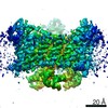
- PDB-6sy7: Structure of Trypanosome Brucei Phosphofructokinase in complex wi... -
+ Open data

- Basic information
| Entry | Database: PDB / ID: 6sy7 | ||||||
|---|---|---|---|---|---|---|---|
| Title | Structure of Trypanosome Brucei Phosphofructokinase in complex with AMP. | ||||||
 Components Components | ATP-dependent 6-phosphofructokinase | ||||||
 Keywords Keywords | TRANSFERASE / Glycolysis / AMP / Allostery | ||||||
| Function / homology |  Function and homology information Function and homology information6-phosphofructokinase / 6-phosphofructokinase activity / glycosome / fructose 6-phosphate metabolic process / phosphate ion binding / glycolytic process / ATP binding / metal ion binding Similarity search - Function | ||||||
| Biological species |  | ||||||
| Method |  X-RAY DIFFRACTION / SYNCHROTRON / MOLECULAR REPLACEMENT / Resolution: 2.75 Å X-RAY DIFFRACTION / SYNCHROTRON / MOLECULAR REPLACEMENT / Resolution: 2.75 Å | ||||||
 Authors Authors | McNae, I.W. / Vasquez-Valdivieso, M.G. / Walkinshaw, M.D. | ||||||
 Citation Citation | Journal: Febs J. / Year: 2020 Title: Kinetic and structural studies of Trypanosoma and Leishmania phosphofructokinases show evolutionary divergence and identify AMP as a switch regulating glycolysis versus gluconeogenesis. Authors: Fernandes, P.M. / Kinkead, J. / McNae, I.W. / Vasquez-Valdivieso, M. / Wear, M.A. / Michels, P.A.M. / Walkinshaw, M.D. | ||||||
| History |
|
- Structure visualization
| Structure viewer | Molecule: MolmilJmol/JSmol |
|---|
- Downloads & links
-Download
| PDBx/mmCIF format | 6sy7.cif.gz | 707.2 KB | Display | PDBx/mmCIF format |
|---|---|---|---|---|
| PDB format | pdb6sy7.ent.gz | 576.8 KB | Display | PDB format |
| PDBx/mmJSON format | 6sy7.json.gz | Tree view | PDBx/mmJSON format | |
| Others |  Other downloads Other downloads |
-Validation report
| Arichive directory | https://data.pdbj.org/pub/pdb/validation_reports/sy/6sy7ftp://data.pdbj.org/pub/pdb/validation_reports/sy/6sy7 | HTTPS FTP |
|---|
-Related structure data
| Related structure data |  3f5mS S: Starting model for refinement |
|---|---|
| Similar structure data |


-Links
 PDBj
PDBj


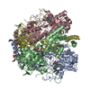
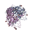
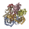
- Assembly
| Deposited unit | 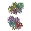
| ||||||||||||
|---|---|---|---|---|---|---|---|---|---|---|---|---|---|
| 1 | 
| ||||||||||||
| 2 | 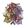
| ||||||||||||
| Unit cell |
|
-Components
| #1: Protein | Mass: 53593.348 Da / Num. of mol.: 8 Source method: isolated from a genetically manipulated source Source: (gene. exp.)  #2: Chemical | ChemComp-BNZ / 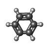  Mass: 78.112 Da / Num. of mol.: 8 / Source method: obtained synthetically / Formula: C6H6 Mass: 78.112 Da / Num. of mol.: 8 / Source method: obtained synthetically / Formula: C6H6#3: Chemical | ChemComp-GOL /   Mass: 92.094 Da / Num. of mol.: 8 / Source method: obtained synthetically / Formula: C3H8O3 Mass: 92.094 Da / Num. of mol.: 8 / Source method: obtained synthetically / Formula: C3H8O3#4: Chemical | ChemComp-AMP /   Mass: 347.221 Da / Num. of mol.: 8 / Source method: obtained synthetically / Formula: C10H14N5O7P / Feature type: SUBJECT OF INVESTIGATION / Comment: AMP*YM Mass: 347.221 Da / Num. of mol.: 8 / Source method: obtained synthetically / Formula: C10H14N5O7P / Feature type: SUBJECT OF INVESTIGATION / Comment: AMP*YM#5: Water | ChemComp-HOH / |  Mass: 18.015 Da / Num. of mol.: 640 / Source method: isolated from a natural source / Formula: H2O Mass: 18.015 Da / Num. of mol.: 640 / Source method: isolated from a natural source / Formula: H2OHas ligand of interest | Y | |
|---|
-Experimental details
-Experiment
| Experiment | Method: X-RAY DIFFRACTION / Number of used crystals: 1 |
|---|
- Sample preparation
| Crystal | Density Matthews: 2.51 Å3/Da / Density % sol: 50.98 % |
|---|---|
| Crystal grow | Temperature: 291 K / Method: vapor diffusion, hanging drop / Details: PEG 8000, sodium cacodylate |
-Data collection
| Diffraction | Mean temperature: 100 K / Serial crystal experiment: N |
|---|---|
| Diffraction source | Source: SYNCHROTRON / Site: Diamond  / Beamline: I04-1 / Wavelength: 0.9159 Å / Beamline: I04-1 / Wavelength: 0.9159 Å |
| Detector | Type: DECTRIS PILATUS3 6M / Detector: PIXEL / Date: Nov 15, 2011 |
| Radiation | Protocol: SINGLE WAVELENGTH / Monochromatic (M) / Laue (L): M / Scattering type: x-ray |
| Radiation wavelength | Wavelength: 0.9159 Å / Relative weight: 1 |
| Reflection | Resolution: 2.75→39.81 Å / Num. obs: 111370 / % possible obs: 99 % / Redundancy: 3.8 % / CC1/2: 0.997 / Rmerge(I) obs: 0.075 / Rrim(I) all: 0.1 / Rsym value: 0.05 / Net I/σ(I): 9.6 |
| Reflection shell | Resolution: 2.75→2.848 Å / Rmerge(I) obs: 0.604 / Mean I/σ(I) obs: 1.5 / Num. unique obs: 5467 / CC1/2: 0.694 / Rpim(I) all: 0.398 / Rrim(I) all: 0.808 |
- Processing
| Software |
| ||||||||||||||||||||||||
|---|---|---|---|---|---|---|---|---|---|---|---|---|---|---|---|---|---|---|---|---|---|---|---|---|---|
| Refinement | Method to determine structure: MOLECULAR REPLACEMENT Starting model: 3F5M Resolution: 2.75→39.81 Å / Cross valid method: FREE R-VALUE /
| ||||||||||||||||||||||||
| Displacement parameters | Biso mean: 34.85 Å2 | ||||||||||||||||||||||||
| Refinement step | Cycle: LAST / Resolution: 2.75→39.81 Å
| ||||||||||||||||||||||||
| Refine LS restraints |
|
