 Movie
Movie Controller

[English] 日本語
 Yorodumi
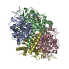
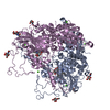
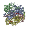
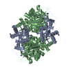
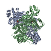
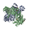
- PDB-6qu4: Crystal Structure of Phosphofructokinase from Trypanosoma brucei ... -
+ Open data

- Basic information
| Entry | Database: PDB / ID: 6qu4 | ||||||
|---|---|---|---|---|---|---|---|
| Title | Crystal Structure of Phosphofructokinase from Trypanosoma brucei in complex with an allosteric inhibitor ctcb405 | ||||||
 Components Components | ATP-dependent 6-phosphofructokinase | ||||||
 Keywords Keywords | TRANSFERASE / Glycolysis / Inhibitor / Allostery | ||||||
| Function / homology |  Function and homology information Function and homology information6-phosphofructokinase / 6-phosphofructokinase activity / glycosome / fructose 6-phosphate metabolic process / phosphate ion binding / glycolytic process / ATP binding / metal ion binding Similarity search - Function | ||||||
| Biological species |  | ||||||
| Method |  X-RAY DIFFRACTION / SYNCHROTRON / MOLECULAR REPLACEMENT / Resolution: 2.75 Å X-RAY DIFFRACTION / SYNCHROTRON / MOLECULAR REPLACEMENT / Resolution: 2.75 Å | ||||||
 Authors Authors | McNae, I.W. / Dornan, J. / Walkinshaw, M.D. | ||||||
| Funding support |  United Kingdom, 1items United Kingdom, 1items
| ||||||
 Citation Citation | Journal: Nat Commun / Year: 2021 Title: Fast acting allosteric phosphofructokinase inhibitors block trypanosome glycolysis and cure acute African trypanosomiasis in mice. Authors: McNae, I.W. / Kinkead, J. / Malik, D. / Yen, L.H. / Walker, M.K. / Swain, C. / Webster, S.P. / Gray, N. / Fernandes, P.M. / Myburgh, E. / Blackburn, E.A. / Ritchie, R. / Austin, C. / Wear, M. ...Authors: McNae, I.W. / Kinkead, J. / Malik, D. / Yen, L.H. / Walker, M.K. / Swain, C. / Webster, S.P. / Gray, N. / Fernandes, P.M. / Myburgh, E. / Blackburn, E.A. / Ritchie, R. / Austin, C. / Wear, M.A. / Highton, A.J. / Keats, A.J. / Vong, A. / Dornan, J. / Mottram, J.C. / Michels, P.A.M. / Pettit, S. / Walkinshaw, M.D. | ||||||
| History |
|
- Structure visualization
| Structure viewer | Molecule: MolmilJmol/JSmol |
|---|
- Downloads & links
-Download
| PDBx/mmCIF format | 6qu4.cif.gz | 702.8 KB | Display | PDBx/mmCIF format |
|---|---|---|---|---|
| PDB format | pdb6qu4.ent.gz | 585.9 KB | Display | PDB format |
| PDBx/mmJSON format | 6qu4.json.gz | Tree view | PDBx/mmJSON format | |
| Others |  Other downloads Other downloads |
-Validation report
| Arichive directory | https://data.pdbj.org/pub/pdb/validation_reports/qu/6qu4ftp://data.pdbj.org/pub/pdb/validation_reports/qu/6qu4 | HTTPS FTP |
|---|
-Related structure data
| Related structure data |  6qu3C  6qu5C  3f5mS S: Starting model for refinement C: citing same article ( |
|---|---|
| Similar structure data |


-Links
 PDBj
PDBj
- Assembly
| Deposited unit | 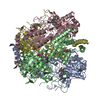
| ||||||||||||||||||||||||||||||||||||||||||||||||||||||||||||||||||||||||||||||||||||||||||||||||||||||||||||||||||||||||||||||||||||||||||||||||||||||||||||||||||||||||||||||||
|---|---|---|---|---|---|---|---|---|---|---|---|---|---|---|---|---|---|---|---|---|---|---|---|---|---|---|---|---|---|---|---|---|---|---|---|---|---|---|---|---|---|---|---|---|---|---|---|---|---|---|---|---|---|---|---|---|---|---|---|---|---|---|---|---|---|---|---|---|---|---|---|---|---|---|---|---|---|---|---|---|---|---|---|---|---|---|---|---|---|---|---|---|---|---|---|---|---|---|---|---|---|---|---|---|---|---|---|---|---|---|---|---|---|---|---|---|---|---|---|---|---|---|---|---|---|---|---|---|---|---|---|---|---|---|---|---|---|---|---|---|---|---|---|---|---|---|---|---|---|---|---|---|---|---|---|---|---|---|---|---|---|---|---|---|---|---|---|---|---|---|---|---|---|---|---|---|---|
| 1 |
| ||||||||||||||||||||||||||||||||||||||||||||||||||||||||||||||||||||||||||||||||||||||||||||||||||||||||||||||||||||||||||||||||||||||||||||||||||||||||||||||||||||||||||||||||
| Unit cell |
| ||||||||||||||||||||||||||||||||||||||||||||||||||||||||||||||||||||||||||||||||||||||||||||||||||||||||||||||||||||||||||||||||||||||||||||||||||||||||||||||||||||||||||||||||
| Noncrystallographic symmetry (NCS) | NCS domain:
NCS domain segments: Component-ID: _ / Refine code: _
NCS ensembles :
|
-Components
| #1: Protein | Mass: 55764.707 Da / Num. of mol.: 4 Source method: isolated from a genetically manipulated source Source: (gene. exp.)  #2: Chemical | ChemComp-JJ2 / 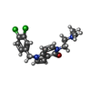  Mass: 364.269 Da / Num. of mol.: 4 / Source method: obtained synthetically / Formula: C18H19Cl2N3O Mass: 364.269 Da / Num. of mol.: 4 / Source method: obtained synthetically / Formula: C18H19Cl2N3O#3: Chemical | ChemComp-CIT /   Mass: 192.124 Da / Num. of mol.: 4 / Source method: obtained synthetically / Formula: C6H8O7 Mass: 192.124 Da / Num. of mol.: 4 / Source method: obtained synthetically / Formula: C6H8O7#4: Chemical | ChemComp-GOL /   Mass: 92.094 Da / Num. of mol.: 4 / Source method: obtained synthetically / Formula: C3H8O3 Mass: 92.094 Da / Num. of mol.: 4 / Source method: obtained synthetically / Formula: C3H8O3#5: Water | ChemComp-HOH / |  Mass: 18.015 Da / Num. of mol.: 72 / Source method: isolated from a natural source / Formula: H2O Mass: 18.015 Da / Num. of mol.: 72 / Source method: isolated from a natural source / Formula: H2O |
|---|
-Experimental details
-Experiment
| Experiment | Method: X-RAY DIFFRACTION / Number of used crystals: 1 |
|---|
- Sample preparation
| Crystal | Density Matthews: 2.63 Å3/Da / Density % sol: 53.15 % |
|---|---|
| Crystal grow | Temperature: 290 K / Method: vapor diffusion, hanging drop Details: 0.1M carboxylic acids (sodium formate, ammonium acetate, sodium citrate tribasic dihydrate, sodium 154 potassium tartrate tetrahydrate and sodium oxamate), buffer system 1 (0.1 M imidazole; ...Details: 0.1M carboxylic acids (sodium formate, ammonium acetate, sodium citrate tribasic dihydrate, sodium 154 potassium tartrate tetrahydrate and sodium oxamate), buffer system 1 (0.1 M imidazole; MES 155 monohydrate (acid)) and precipitant mix 3 (40% v/v glycerol; 20% w/v PEG 4000) |
-Data collection
| Diffraction | Mean temperature: 100 K / Serial crystal experiment: N |
|---|---|
| Diffraction source | Source: SYNCHROTRON / Site: Diamond / Beamline: I24 / Wavelength: 0.9685 Å |
| Detector | Type: DECTRIS PILATUS3 6M / Detector: PIXEL / Date: Dec 7, 2016 |
| Radiation | Protocol: SINGLE WAVELENGTH / Monochromatic (M) / Laue (L): M / Scattering type: x-ray |
| Radiation wavelength | Wavelength: 0.9685 Å / Relative weight: 1 |
| Reflection | Resolution: 2.75→48.02 Å / Num. obs: 50992 / % possible obs: 92.98 % / Redundancy: 4 % / Net I/σ(I): 9.5 |
| Reflection shell | Resolution: 2.75→2.848 Å |
- Processing
| Software |
| ||||||||||||||||||||||||||||||||||||||||||||||||||||||||||||||||||||||||||||||||||||||||||||||||||||||||||||||||||||||||||||||||||||||||||||||||||||||||||||||||||||||||||||||||||||||
|---|---|---|---|---|---|---|---|---|---|---|---|---|---|---|---|---|---|---|---|---|---|---|---|---|---|---|---|---|---|---|---|---|---|---|---|---|---|---|---|---|---|---|---|---|---|---|---|---|---|---|---|---|---|---|---|---|---|---|---|---|---|---|---|---|---|---|---|---|---|---|---|---|---|---|---|---|---|---|---|---|---|---|---|---|---|---|---|---|---|---|---|---|---|---|---|---|---|---|---|---|---|---|---|---|---|---|---|---|---|---|---|---|---|---|---|---|---|---|---|---|---|---|---|---|---|---|---|---|---|---|---|---|---|---|---|---|---|---|---|---|---|---|---|---|---|---|---|---|---|---|---|---|---|---|---|---|---|---|---|---|---|---|---|---|---|---|---|---|---|---|---|---|---|---|---|---|---|---|---|---|---|---|---|
| Refinement | Method to determine structure: MOLECULAR REPLACEMENT Starting model: 3f5m Resolution: 2.75→48.02 Å / Cor.coef. Fo:Fc: 0.926 / Cor.coef. Fo:Fc free: 0.899 / SU B: 42.894 / SU ML: 0.368 / Cross valid method: THROUGHOUT / ESU R Free: 0.397 / Details: HYDROGENS HAVE BEEN ADDED IN THE RIDING POSITIONS
| ||||||||||||||||||||||||||||||||||||||||||||||||||||||||||||||||||||||||||||||||||||||||||||||||||||||||||||||||||||||||||||||||||||||||||||||||||||||||||||||||||||||||||||||||||||||
| Solvent computation | Ion probe radii: 0.7 Å / Shrinkage radii: 0.7 Å / VDW probe radii: 1.1 Å | ||||||||||||||||||||||||||||||||||||||||||||||||||||||||||||||||||||||||||||||||||||||||||||||||||||||||||||||||||||||||||||||||||||||||||||||||||||||||||||||||||||||||||||||||||||||
| Displacement parameters | Biso mean: 62.15 Å2
| ||||||||||||||||||||||||||||||||||||||||||||||||||||||||||||||||||||||||||||||||||||||||||||||||||||||||||||||||||||||||||||||||||||||||||||||||||||||||||||||||||||||||||||||||||||||
| Refinement step | Cycle: LAST / Resolution: 2.75→48.02 Å
| ||||||||||||||||||||||||||||||||||||||||||||||||||||||||||||||||||||||||||||||||||||||||||||||||||||||||||||||||||||||||||||||||||||||||||||||||||||||||||||||||||||||||||||||||||||||
| Refine LS restraints |
|