 Movie
Movie Controller

[English] 日本語
 Yorodumi
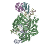
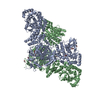
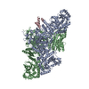
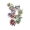
- PDB-6swy: Structure of active GID E3 ubiquitin ligase complex minus Gid2 an... -
+ Open data

- Basic information
| Entry | Database: PDB / ID: 6swy | |||||||||
|---|---|---|---|---|---|---|---|---|---|---|
| Title | Structure of active GID E3 ubiquitin ligase complex minus Gid2 and delta Gid9 RING domain | |||||||||
 Components Components |
| |||||||||
 Keywords Keywords | LIGASE / Suppressed / Suppreseed | |||||||||
| Function / homology |  Function and homology information Function and homology information: / protein catabolic process in the vacuole / GID complex / ubiquitin-dependent protein catabolic process via the N-end rule pathway / ascospore formation / traversing start control point of mitotic cell cycle / regulation of nitrogen utilization / protein targeting to vacuole / vacuole / negative regulation of gluconeogenesis ...: / protein catabolic process in the vacuole / GID complex / ubiquitin-dependent protein catabolic process via the N-end rule pathway / ascospore formation / traversing start control point of mitotic cell cycle / regulation of nitrogen utilization / protein targeting to vacuole / vacuole / negative regulation of gluconeogenesis / Neutrophil degranulation / cytoskeleton organization / cytoplasmic vesicle membrane / RING-type E3 ubiquitin transferase / ubiquitin protein ligase activity / cytoplasmic vesicle / proteasome-mediated ubiquitin-dependent protein catabolic process / protein ubiquitination / negative regulation of apoptotic process / zinc ion binding / nucleus / cytosol / cytoplasm Similarity search - Function | |||||||||
| Biological species |  | |||||||||
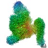
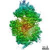
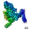
| Method | ELECTRON MICROSCOPY / single particle reconstruction / cryo EM / Resolution: 3.2 Å | |||||||||
 Authors Authors | Qiao, S. / Prabu, J.R. / Schulman, B.A. | |||||||||
| Funding support |  Germany, 1items Germany, 1items
| |||||||||
 Citation Citation | Journal: Mol Cell / Year: 2020 Title: Interconversion between Anticipatory and Active GID E3 Ubiquitin Ligase Conformations via Metabolically Driven Substrate Receptor Assembly. Authors: Shuai Qiao / Christine R Langlois / Jakub Chrustowicz / Dawafuti Sherpa / Ozge Karayel / Fynn M Hansen / Viola Beier / Susanne von Gronau / Daniel Bollschweiler / Tillman Schäfer / Arno F ...Authors: Shuai Qiao / Christine R Langlois / Jakub Chrustowicz / Dawafuti Sherpa / Ozge Karayel / Fynn M Hansen / Viola Beier / Susanne von Gronau / Daniel Bollschweiler / Tillman Schäfer / Arno F Alpi / Matthias Mann / J Rajan Prabu / Brenda A Schulman / Abstract: Cells respond to environmental changes by toggling metabolic pathways, preparing for homeostasis, and anticipating future stresses. For example, in Saccharomyces cerevisiae, carbon stress-induced ...Cells respond to environmental changes by toggling metabolic pathways, preparing for homeostasis, and anticipating future stresses. For example, in Saccharomyces cerevisiae, carbon stress-induced gluconeogenesis is terminated upon glucose availability, a process that involves the multiprotein E3 ligase GID recruiting N termini and catalyzing ubiquitylation of gluconeogenic enzymes. Here, genetics, biochemistry, and cryoelectron microscopy define molecular underpinnings of glucose-induced degradation. Unexpectedly, carbon stress induces an inactive anticipatory complex (GID), which awaits a glucose-induced substrate receptor to form the active GID. Meanwhile, other environmental perturbations elicit production of an alternative substrate receptor assembling into a related E3 ligase complex. The intricate structure of GID enables anticipating and ultimately binding various N-degron-targeting (i.e., "N-end rule") substrate receptors, while the GID E3 forms a clamp-like structure juxtaposing substrate lysines with the ubiquitylation active site. The data reveal evolutionarily conserved GID complexes as a family of multisubunit E3 ubiquitin ligases responsive to extracellular stimuli. | |||||||||
| History |
|
- Structure visualization
| Movie |
Movie viewer |
|---|---|
| Structure viewer | Molecule: MolmilJmol/JSmol |
- Downloads & links
-Download
| PDBx/mmCIF format | 6swy.cif.gz | 387.5 KB | Display | PDBx/mmCIF format |
|---|---|---|---|---|
| PDB format | pdb6swy.ent.gz | 296 KB | Display | PDB format |
| PDBx/mmJSON format | 6swy.json.gz | Tree view | PDBx/mmJSON format | |
| Others |  Other downloads Other downloads |
-Validation report
| Arichive directory | https://data.pdbj.org/pub/pdb/validation_reports/sw/6swyftp://data.pdbj.org/pub/pdb/validation_reports/sw/6swy | HTTPS FTP |
|---|
-Related structure data
| Related structure data | 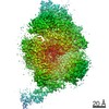 10333MC C: citing same article ( M: map data used to model this data |
|---|---|
| Similar structure data |


-Links
 PDBj
PDBj


- Assembly
| Deposited unit | 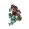
|
|---|---|
| 1 |
|
-Components
| #1: Protein | Mass: 105658.203 Da / Num. of mol.: 1 Source method: isolated from a genetically manipulated source Source: (gene. exp.) Strain: ATCC 204508 / S288c / Gene: VID28, GID5, YIL017C Production host:  Spodoptera aff. frugiperda 1 BOLD-2017 (butterflies/moths) Spodoptera aff. frugiperda 1 BOLD-2017 (butterflies/moths)References: UniProt: P40547 |
|---|---|
| #2: Protein | Mass: 51789.152 Da / Num. of mol.: 1 Source method: isolated from a genetically manipulated source Source: (gene. exp.) Production host: Spodoptera aff. frugiperda 1 BOLD-2017 (butterflies/moths)References: UniProt: P40208 |
| #3: Protein | Mass: 117308.906 Da / Num. of mol.: 1 Source method: isolated from a genetically manipulated source Source: (gene. exp.) Gene: VID30, GID1, YGL227W Production host: Spodoptera aff. frugiperda 1 BOLD-2017 (butterflies/moths)References: UniProt: P53076 |
| #4: Protein | Mass: 41291.934 Da / Num. of mol.: 1 Source method: isolated from a genetically manipulated source Source: (gene. exp.) Production host: Spodoptera aff. frugiperda 1 BOLD-2017 (butterflies/moths)References: UniProt: P38263 |
| #5: Protein | Mass: 54432.879 Da / Num. of mol.: 1 Source method: isolated from a genetically manipulated source Source: (gene. exp.) Gene: FYV10, GID9, YIL097W Production host: Spodoptera aff. frugiperda 1 BOLD-2017 (butterflies/moths)References: UniProt: P40492, RING-type E3 ubiquitin transferase |
| Compound details | For residue numbers above 3000 could potentially correspond to the indicated chain, although their ...For residue numbers above 3000 could potentially correspond to the indicated chain, although their precise positions and identities are ambiguous. |
-Experimental details
-Experiment
| Experiment | Method: ELECTRON MICROSCOPY |
|---|---|
| EM experiment | Aggregation state: PARTICLE / 3D reconstruction method: single particle reconstruction |
- Sample preparation
| Component | Name: GIDSR4 minus Gid2/delta Gid9RING / Type: COMPLEX / Entity ID: all / Source: RECOMBINANT |
|---|---|
| Source (natural) | Organism: |
| Source (recombinant) | Organism: Spodoptera aff. frugiperda 1 BOLD-2017 (butterflies/moths) |
| Buffer solution | pH: 6.5 |
| Specimen | Conc.: 0.25 mg/ml / Embedding applied: NO / Shadowing applied: NO / Staining applied: NO / Vitrification applied: YES |
| Vitrification | Cryogen name: ETHANE |
- Electron microscopy imaging
| Experimental equipment |  Model: Titan Krios / Image courtesy: FEI Company |
|---|---|
| Microscopy | Model: FEI TITAN KRIOS |
| Electron gun | Electron source:  FIELD EMISSION GUN / Accelerating voltage: 300 kV / Illumination mode: FLOOD BEAM FIELD EMISSION GUN / Accelerating voltage: 300 kV / Illumination mode: FLOOD BEAM |
| Electron lens | Mode: BRIGHT FIELD |
| Image recording | Electron dose: 54.48 e/Å2 / Film or detector model: GATAN K3 BIOQUANTUM (6k x 4k) |
- Processing
| Software | Name: PHENIX / Version: 1.18.2_3874: / Classification: refinement |
|---|---|
| EM software | Name: RELION / Version: 3 / Category: 3D reconstruction |
| CTF correction | Type: PHASE FLIPPING AND AMPLITUDE CORRECTION |
| Symmetry | Point symmetry: C1 (asymmetric) |
| 3D reconstruction | Resolution: 3.2 Å / Resolution method: FSC 0.143 CUT-OFF / Num. of particles: 122248 / Symmetry type: POINT |
