 Movie
Movie Controller

[English] 日本語
 Yorodumi
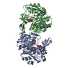
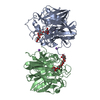
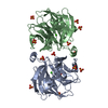
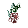
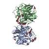
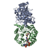
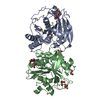
- PDB-6su9: Crystal structure of Plasmodium falciparum PdxK with ligands AMP-... -
+ Open data

- Basic information
| Entry | Database: PDB / ID: 6su9 | ||||||
|---|---|---|---|---|---|---|---|
| Title | Crystal structure of Plasmodium falciparum PdxK with ligands AMP-PNP and PL | ||||||
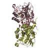
 Components Components | Pyridoxal kinase | ||||||
 Keywords Keywords | TRANSFERASE / Crystal structure of Plasmodium falciparum PdxK with ligands AMP-PNP and PL. | ||||||
| Function / homology |  Function and homology information Function and homology informationpyridoxamine binding / pyridoxine binding / Vitamin B6 activation to pyridoxal phosphate / vitamin B6 biosynthetic process / pyridoxal binding / pyridoxine metabolic process / pyridoxal 5'-phosphate salvage / pyridoxal kinase / pyridoxal kinase activity / response to singlet oxygen ...pyridoxamine binding / pyridoxine binding / Vitamin B6 activation to pyridoxal phosphate / vitamin B6 biosynthetic process / pyridoxal binding / pyridoxine metabolic process / pyridoxal 5'-phosphate salvage / pyridoxal kinase / pyridoxal kinase activity / response to singlet oxygen / Neutrophil degranulation / phosphorylation / ATP binding / metal ion binding / cytosol Similarity search - Function | ||||||
| Biological species |  | ||||||
| Method |  X-RAY DIFFRACTION / SYNCHROTRON / MOLECULAR REPLACEMENT / Resolution: 2.15 Å X-RAY DIFFRACTION / SYNCHROTRON / MOLECULAR REPLACEMENT / Resolution: 2.15 Å | ||||||
 Authors Authors | Groves, M.R. / Gao, K. / Wang, W. | ||||||
 Citation Citation | Journal: Crystals / Year: 2019 Title: Crystal structure of Plasmodium falciparum PdxK with ligands AMP-PNP and PL. Authors: Gao, K. / Wang, W. / Groves, M.R. | ||||||
| History |
|
- Structure visualization
| Structure viewer | Molecule: MolmilJmol/JSmol |
|---|
- Downloads & links
-Download
| PDBx/mmCIF format | 6su9.cif.gz | 268.2 KB | Display | PDBx/mmCIF format |
|---|---|---|---|---|
| PDB format | pdb6su9.ent.gz | 212.1 KB | Display | PDB format |
| PDBx/mmJSON format | 6su9.json.gz | Tree view | PDBx/mmJSON format | |
| Others |  Other downloads Other downloads |
-Validation report
| Arichive directory | https://data.pdbj.org/pub/pdb/validation_reports/su/6su9ftp://data.pdbj.org/pub/pdb/validation_reports/su/6su9 | HTTPS FTP |
|---|
-Related structure data
| Related structure data | 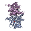 1rfuS S: Starting model for refinement |
|---|---|
| Similar structure data |


-Links
 PDBj
PDBj


- Assembly
| Deposited unit | 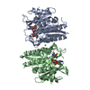
| ||||||||
|---|---|---|---|---|---|---|---|---|---|
| 1 |
| ||||||||
| Unit cell |
|
-Components
| #1: Protein | Mass: 57277.742 Da / Num. of mol.: 2 Source method: isolated from a genetically manipulated source Source: (gene. exp.) Strain: isolate 3D7 / Gene: PF3D7_0616000 / Production host:  #2: Chemical |   Mass: 506.196 Da / Num. of mol.: 2 / Source method: obtained synthetically / Formula: C10H17N6O12P3 / Comment: AMP-PNP, energy-carrying molecule analogue*YM Mass: 506.196 Da / Num. of mol.: 2 / Source method: obtained synthetically / Formula: C10H17N6O12P3 / Comment: AMP-PNP, energy-carrying molecule analogue*YM#3: Chemical | 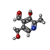  Mass: 167.162 Da / Num. of mol.: 2 / Source method: obtained synthetically / Formula: C8H9NO3 Mass: 167.162 Da / Num. of mol.: 2 / Source method: obtained synthetically / Formula: C8H9NO3#4: Chemical |   Mass: 24.305 Da / Num. of mol.: 3 / Source method: obtained synthetically / Formula: Mg Mass: 24.305 Da / Num. of mol.: 3 / Source method: obtained synthetically / Formula: Mg#5: Water | ChemComp-HOH / |  Mass: 18.015 Da / Num. of mol.: 69 / Source method: isolated from a natural source / Formula: H2O Mass: 18.015 Da / Num. of mol.: 69 / Source method: isolated from a natural source / Formula: H2OHas ligand of interest | N | |
|---|
-Experimental details
-Experiment
| Experiment | Method: X-RAY DIFFRACTION / Number of used crystals: 1 |
|---|
- Sample preparation
| Crystal | Density Matthews: 2.25 Å3/Da / Density % sol: 56.01 % |
|---|---|
| Crystal grow | Temperature: 293 K / Method: vapor diffusion, hanging drop / pH: 7.8 Details: 0.1 M HEPES pH 7.75, 0.2 M CaCl2, 31%(v/v) PEG400, 5%(v/v) glycerol PH range: 7.0-8.0 |
-Data collection
| Diffraction | Mean temperature: 100 K / Serial crystal experiment: N |
|---|---|
| Diffraction source | Source: SYNCHROTRON / Site: PETRA III, EMBL c/o DESY  / Beamline: P13 (MX1) / Wavelength: 0.97 Å / Beamline: P13 (MX1) / Wavelength: 0.97 Å |
| Detector | Type: DECTRIS PILATUS3 S 6M / Detector: PIXEL / Date: Jul 8, 2014 |
| Radiation | Protocol: SINGLE WAVELENGTH / Monochromatic (M) / Laue (L): M / Scattering type: x-ray |
| Radiation wavelength | Wavelength: 0.97 Å / Relative weight: 1 |
| Reflection | Resolution: 2.15→19.33 Å / Num. obs: 32284 / % possible obs: 97.87 % / Redundancy: 2.55 % / Rmerge(I) obs: 0.042 / Net I/σ(I): 2 |
| Reflection shell | Resolution: 2.15→2.227 Å / Rmerge(I) obs: 0.042 / Num. unique obs: 3159 / % possible all: 97.17 |
- Processing
| Software |
| ||||||||||||||||||||||||||||||||||||||||||||||||||||||||||||||||||||||||||||||
|---|---|---|---|---|---|---|---|---|---|---|---|---|---|---|---|---|---|---|---|---|---|---|---|---|---|---|---|---|---|---|---|---|---|---|---|---|---|---|---|---|---|---|---|---|---|---|---|---|---|---|---|---|---|---|---|---|---|---|---|---|---|---|---|---|---|---|---|---|---|---|---|---|---|---|---|---|---|---|---|
| Refinement | Method to determine structure: MOLECULAR REPLACEMENT Starting model: 1RFU Resolution: 2.15→19.327 Å / SU ML: 0.28 / Cross valid method: THROUGHOUT / σ(F): 1.34 / Phase error: 31.93
| ||||||||||||||||||||||||||||||||||||||||||||||||||||||||||||||||||||||||||||||
| Solvent computation | Shrinkage radii: 0.9 Å / VDW probe radii: 1.11 Å | ||||||||||||||||||||||||||||||||||||||||||||||||||||||||||||||||||||||||||||||
| Displacement parameters | Biso max: 131.43 Å2 / Biso mean: 59.7016 Å2 / Biso min: 20.85 Å2 | ||||||||||||||||||||||||||||||||||||||||||||||||||||||||||||||||||||||||||||||
| Refinement step | Cycle: final / Resolution: 2.15→19.327 Å
| ||||||||||||||||||||||||||||||||||||||||||||||||||||||||||||||||||||||||||||||
| Refine LS restraints |
| ||||||||||||||||||||||||||||||||||||||||||||||||||||||||||||||||||||||||||||||
| LS refinement shell | Refine-ID: X-RAY DIFFRACTION / Rfactor Rfree error: 0
| ||||||||||||||||||||||||||||||||||||||||||||||||||||||||||||||||||||||||||||||
| Refinement TLS params. | Method: refined / Origin x: 57.9389 Å / Origin y: -0.6275 Å / Origin z: 23.2296 Å
| ||||||||||||||||||||||||||||||||||||||||||||||||||||||||||||||||||||||||||||||
| Refinement TLS group |
|
