 Movie
Movie Controller


+ Open data

- Basic information
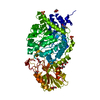
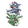
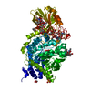
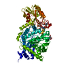
| Entry | Database: PDB / ID: 1rfu | ||||||
|---|---|---|---|---|---|---|---|
| Title | Crystal structure of pyridoxal kinase complexed with ADP and PLP | ||||||
 Components Components | pyridoxal kinase | ||||||
 Keywords Keywords | TRANSFERASE | ||||||
| Function / homology |  Function and homology information Function and homology informationpyridoxal 5'-phosphate salvage / pyridoxal kinase / pyridoxal kinase activity / ATP binding / metal ion binding / cytosol Similarity search - Function | ||||||
| Biological species |  | ||||||
| Method |  X-RAY DIFFRACTION / MOLECULAR REPLACEMENT / Resolution: 2.8 Å X-RAY DIFFRACTION / MOLECULAR REPLACEMENT / Resolution: 2.8 Å | ||||||
 Authors Authors | Liang, D.-C. / Jiang, T. / Li, M.-H. | ||||||
 Citation Citation | Journal: J.BIOL.CHEM. / Year: 2004 Title: Conformational changes in the reaction of pyridoxal kinase Authors: Li, M.-H. / Kwok, F. / Chang, W.-R. / Liu, S.-Q. / Lo, S.C.L. / Zhang, J.-P. / Jiang, T. / Liang, D.-C. #1: Journal: J.Biol.Chem. / Year: 2002Title: Crystal Structure of Brain Pyridoxal Kinase, a Novel Member of the Ribokinase Superfamily Authors: Li, M.-H. / Kwok, F. / Chang, W.-R. / Lau, C.-K. / Zhang, J.-P. / Lo, S.C.L. / Jiang, T. / Liang, D.-C. #2: Journal: Acta Crystallogr.,Sect.D / Year: 2002 Title: Crystallization and preliminary crystallographic studies of pyridoxal kinase from sheep brain Authors: Li, M.-H. / Kwok, F. / An, X.-M. / Chang, W.-R. / Lau, C.-K. / Zhang, J.-P. / Liu, S.-Q. / Leung, Y.-C. / Jiang, T. / Liang, D.-C. | ||||||
| History |
|
- Structure visualization
| Structure viewer | Molecule: MolmilJmol/JSmol |
|---|
- Downloads & links
-Download
| PDBx/mmCIF format | 1rfu.cif.gz | 492.3 KB | Display | PDBx/mmCIF format |
|---|---|---|---|---|
| PDB format | pdb1rfu.ent.gz | 405.2 KB | Display | PDB format |
| PDBx/mmJSON format | 1rfu.json.gz | Tree view | PDBx/mmJSON format | |
| Others |  Other downloads Other downloads |
-Validation report
| Arichive directory | https://data.pdbj.org/pub/pdb/validation_reports/rf/1rfuftp://data.pdbj.org/pub/pdb/validation_reports/rf/1rfu | HTTPS FTP |
|---|
-Related structure data
| Related structure data | 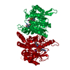 1rftC 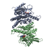 1rfvC 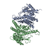 1lhpS S: Starting model for refinement C: citing same article ( |
|---|---|
| Similar structure data |

-Links
 PDBj
PDBj- Assembly
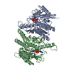
| Deposited unit | 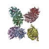
| ||||||||
|---|---|---|---|---|---|---|---|---|---|
| 1 | 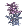
| ||||||||
| 2 | 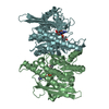
| ||||||||
| 3 | 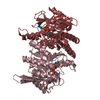
| ||||||||
| 4 | 
| ||||||||
| Unit cell |
| ||||||||
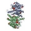
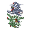
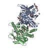
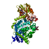
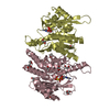
| Details | The biological assembly is a homodimer. The eight monomers in the asymmetric unit form four such homodimers. |
-Components
| #1: Protein | Mass: 34860.879 Da / Num. of mol.: 8 / Source method: isolated from a natural source / Source: (natural) #2: Chemical | ChemComp-ZN /   Mass: 65.409 Da / Num. of mol.: 8 / Source method: obtained synthetically / Formula: Zn Mass: 65.409 Da / Num. of mol.: 8 / Source method: obtained synthetically / Formula: Zn#3: Chemical | ChemComp-PLP /   Mass: 247.142 Da / Num. of mol.: 8 / Source method: obtained synthetically / Formula: C8H10NO6P Mass: 247.142 Da / Num. of mol.: 8 / Source method: obtained synthetically / Formula: C8H10NO6P#4: Chemical | ChemComp-ADP /   Mass: 427.201 Da / Num. of mol.: 8 / Source method: obtained synthetically / Formula: C10H15N5O10P2 / Comment: ADP, energy-carrying molecule*YM Mass: 427.201 Da / Num. of mol.: 8 / Source method: obtained synthetically / Formula: C10H15N5O10P2 / Comment: ADP, energy-carrying molecule*YM#5: Water | ChemComp-HOH / |  Mass: 18.015 Da / Num. of mol.: 267 / Source method: isolated from a natural source / Formula: H2O Mass: 18.015 Da / Num. of mol.: 267 / Source method: isolated from a natural source / Formula: H2O |
|---|
-Experimental details
-Experiment
| Experiment | Method: X-RAY DIFFRACTION / Number of used crystals: 1 |
|---|
- Sample preparation
| Crystal | Density Matthews: 3.04 Å3/Da / Density % sol: 59.25 % |
|---|---|
| Crystal grow | Temperature: 290 K / Method: vapor diffusion, hanging drop / pH: 8.2 Details: ammonium sulphate, potassium phosphate, ADP, PLP, zinc acetate, pH 8.2, VAPOR DIFFUSION, HANGING DROP, temperature 290K |
-Data collection
| Diffraction | Mean temperature: 100 K |
|---|---|
| Diffraction source | Source: SEALED TUBE / Wavelength: 1.5418 Å |
| Detector | Type: MARRESEARCH / Detector: IMAGE PLATE / Date: Aug 12, 2002 |
| Radiation | Monochromator: GRAPHITE / Protocol: SINGLE WAVELENGTH / Monochromatic (M) / Laue (L): M / Scattering type: x-ray |
| Radiation wavelength | Wavelength: 1.5418 Å / Relative weight: 1 |
| Reflection | Resolution: 2.8→20 Å / Num. all: 86300 / Num. obs: 75513 / % possible obs: 87.5 % / Observed criterion σ(F): 0 / Observed criterion σ(I): -3 / Redundancy: 4.8 % / Rmerge(I) obs: 0.087 / Net I/σ(I): 11.4 |
| Reflection shell | Resolution: 2.8→2.86 Å / Rmerge(I) obs: 0.465 / Mean I/σ(I) obs: 2.3 / Num. unique all: 5131 / % possible all: 95.2 |
- Processing
| Software |
| |||||||||||||||||||||||||
|---|---|---|---|---|---|---|---|---|---|---|---|---|---|---|---|---|---|---|---|---|---|---|---|---|---|---|
| Refinement | Method to determine structure: MOLECULAR REPLACEMENT Starting model: PDB ENTRY 1LHP Resolution: 2.8→20 Å / Isotropic thermal model: Isotropic / Cross valid method: THROUGHOUT / σ(F): 0 / σ(I): -3 / Stereochemistry target values: Engh & Huber Details: used least squares procedure for hemihedral twinning, with a twinning operation of "h,-k,-l", and a twinning fraction of 0.5
| |||||||||||||||||||||||||
| Refinement step | Cycle: LAST / Resolution: 2.8→20 Å
| |||||||||||||||||||||||||
| Refine LS restraints |
|
