 Movie
Movie Controller

[English] 日本語
 Yorodumi
- PDB-1j8v: Crystal structure of barley beta-D-glucan glucohydrolase isoenzym... -
+ Open data

- Basic information
| Entry | Database: PDB / ID: 1j8v | |||||||||
|---|---|---|---|---|---|---|---|---|---|---|
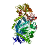
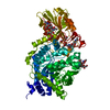
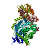
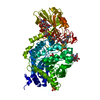
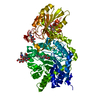
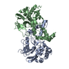
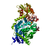
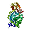
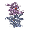
| Title | Crystal structure of barley beta-D-glucan glucohydrolase isoenzyme Exo1 in complex with 4'-nitrophenyl 3I-thiolaminaritrioside | |||||||||
 Components Components | Beta-D-glucan glucohydrolase isoenzyme EXO1 | |||||||||
 Keywords Keywords | HYDROLASE / 2-domain fold / ligand-protein complex | |||||||||
| Function / homology |  Function and homology information Function and homology informationhydrolase activity, hydrolyzing O-glycosyl compounds / carbohydrate metabolic process Similarity search - Function | |||||||||
| Biological species |  | |||||||||
| Method |  X-RAY DIFFRACTION / MOLECULAR REPLACEMENT / Resolution: 2.4 Å X-RAY DIFFRACTION / MOLECULAR REPLACEMENT / Resolution: 2.4 Å | |||||||||
 Authors Authors | Hrmova, M. / De Gori, R. / Smith, B.J. / Fairweather, J.K. / Driguez, H. / Varghese, J.N. / Fincher, G.B. | |||||||||
 Citation Citation | Journal: Plant Cell / Year: 2002 Title: Structural basis for broad substrate specificity in higher plant beta-D-glucan glucohydrolases. Authors: Hrmova, M. / De Gori, R. / Smith, B.J. / Fairweather, J.K. / Driguez, H. / Varghese, J.N. / Fincher, G.B. | |||||||||
| History |
| |||||||||
| Remark 600 | HETEROGEN The only part of the 4'-Nitrophenyl-S-(beta- D-Glucopyranosyl)-(1->3)-(3-thio-beta- D- ...HETEROGEN The only part of the 4'-Nitrophenyl-S-(beta- D-Glucopyranosyl)-(1->3)-(3-thio-beta- D-glucopyranosyl)-(1->3)-beta-D-glucopyranoside ligand seen in the density is the thiolaminaribiosyl portion. The 4'-Nitrophenyl-(beta-D-Glucopyranosyl) moiety is disordered. | |||||||||
| Remark 999 | SEQUENCE The sequence in the GenBank entry might be a sequencing error at residue 345. The electron ...SEQUENCE The sequence in the GenBank entry might be a sequencing error at residue 345. The electron density unambiguously proved the presence of LYS instead of ASN at this residue. |
- Structure visualization
| Structure viewer | Molecule: MolmilJmol/JSmol |
|---|
- Downloads & links
-Download
| PDBx/mmCIF format | 1j8v.cif.gz | 136.9 KB | Display | PDBx/mmCIF format |
|---|---|---|---|---|
| PDB format | pdb1j8v.ent.gz | 104.7 KB | Display | PDB format |
| PDBx/mmJSON format | 1j8v.json.gz | Tree view | PDBx/mmJSON format | |
| Others |  Other downloads Other downloads |
-Validation report
| Arichive directory | https://data.pdbj.org/pub/pdb/validation_reports/j8/1j8vftp://data.pdbj.org/pub/pdb/validation_reports/j8/1j8v | HTTPS FTP |
|---|
-Related structure data
| Related structure data |  1ieqS S: Starting model for refinement |
|---|---|
| Similar structure data |


-Links
 PDBj
PDBj- Assembly
| Deposited unit | 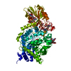
| ||||||||
|---|---|---|---|---|---|---|---|---|---|
| 1 |
| ||||||||
| Unit cell |
| ||||||||
| Details | The biological assembly is a monomer constructed from an (alpha/beta)8 barrel and an (alpha/beta)6 sandwich |
-Components
-Protein , 1 types, 1 molecules A
| #1: Protein | Mass: 65475.617 Da / Num. of mol.: 1 / Source method: isolated from a natural source / Source: (natural) References: GenBank: 4566505, UniProt: Q9XEI3*PLUS, glucan 1,3-beta-glucosidase |
|---|
-Sugars , 3 types, 3 molecules
| #2: Polysaccharide | beta-D-mannopyranose-(1-4)-2-acetamido-2-deoxy-beta-D-glucopyranose-(1-4)-2-acetamido-2-deoxy-beta- ...beta-D-mannopyranose-(1-4)-2-acetamido-2-deoxy-beta-D-glucopyranose-(1-4)-2-acetamido-2-deoxy-beta-D-glucopyranose Source method: isolated from a genetically manipulated source |
|---|---|
| #3: Polysaccharide | 2-acetamido-2-deoxy-beta-D-glucopyranose-(1-2)-alpha-D-mannopyranose-(1-6)-beta-D-mannopyranose-(1- ...2-acetamido-2-deoxy-beta-D-glucopyranose-(1-2)-alpha-D-mannopyranose-(1-6)-beta-D-mannopyranose-(1-4)-2-acetamido-2-deoxy-beta-D-glucopyranose-(1-4)-[alpha-L-fucopyranose-(1-3)]2-acetamido-2-deoxy-beta-D-glucopyranose Source method: isolated from a genetically manipulated source |
| #4: Polysaccharide | 2-acetamido-2-deoxy-beta-D-glucopyranose-(1-4)-2-acetamido-2-deoxy-beta-D-glucopyranose Source method: isolated from a genetically manipulated source |
-Non-polymers , 2 types, 270 molecules 

| #5: Chemical | ChemComp-LAM /  Mass: 641.596 Da / Num. of mol.: 1 / Source method: obtained synthetically / Formula: C24H35NO17S Mass: 641.596 Da / Num. of mol.: 1 / Source method: obtained synthetically / Formula: C24H35NO17S |
|---|---|
| #6: Water | ChemComp-HOH / Mass: 18.015 Da / Num. of mol.: 269 / Source method: isolated from a natural source / Formula: H2O |
-Details
| Has protein modification | Y |
|---|
-Experimental details
-Experiment
| Experiment | Method: X-RAY DIFFRACTION / Number of used crystals: 1 |
|---|
- Sample preparation
| Crystal | Density Matthews: 3.51 Å3/Da / Density % sol: 64.95 % | ||||||||||||||||||||||||||||||||||||||||
|---|---|---|---|---|---|---|---|---|---|---|---|---|---|---|---|---|---|---|---|---|---|---|---|---|---|---|---|---|---|---|---|---|---|---|---|---|---|---|---|---|---|
| Crystal grow | Temperature: 277 K / Method: vapor diffusion, hanging drop / pH: 7 Details: ammonium sulphate, PEG 400, sodium acetate, Hepes-NaOH, pH 7.0, VAPOR DIFFUSION, HANGING DROP, temperature 277K | ||||||||||||||||||||||||||||||||||||||||
| Crystal grow | *PLUS Temperature: 277-279 K / Details: Hrmova, M., (1998) Acta Cryst., D54, 687. | ||||||||||||||||||||||||||||||||||||||||
| Components of the solutions | *PLUS
|
-Data collection
| Diffraction | Mean temperature: 100 K |
|---|---|
| Diffraction source | Source: ROTATING ANODE / Type: MACSCIENCE / Wavelength: 1.5418 Å |
| Detector | Type: RIGAKU RAXIS II / Detector: IMAGE PLATE / Date: Dec 12, 2000 / Details: Focussing mirrors |
| Radiation | Protocol: SINGLE WAVELENGTH / Monochromatic (M) / Laue (L): M / Scattering type: x-ray |
| Radiation wavelength | Wavelength: 1.5418 Å / Relative weight: 1 |
| Reflection | Resolution: 2.4→50 Å / Num. all: 34896 / Num. obs: 34896 / % possible obs: 93.9 % / Observed criterion σ(F): 0 / Observed criterion σ(I): 0 / Redundancy: 5.18 % / Rmerge(I) obs: 0.087 / Net I/σ(I): 7.8 |
| Reflection shell | Resolution: 2.4→50 Å / Rmerge(I) obs: 0.274 / % possible all: 85.5 |
| Reflection | *PLUS Lowest resolution: 50 Å / Num. measured all: 176916 |
| Reflection shell | *PLUS Lowest resolution: 2.46 Å / % possible obs: 85.5 % |
- Processing
| Software |
| |||||||||||||||||||||||||
|---|---|---|---|---|---|---|---|---|---|---|---|---|---|---|---|---|---|---|---|---|---|---|---|---|---|---|
| Refinement | Method to determine structure: MOLECULAR REPLACEMENT Starting model: 1IEQ Resolution: 2.4→10 Å / Isotropic thermal model: Isotropic / Cross valid method: THROUGHOUT / σ(F): 0 / σ(I): 0 / Stereochemistry target values: Engh & Huber
| |||||||||||||||||||||||||
| Refine analyze | Luzzati coordinate error obs: 0.3 Å | |||||||||||||||||||||||||
| Refinement step | Cycle: LAST / Resolution: 2.4→10 Å
| |||||||||||||||||||||||||
| Refine LS restraints |
| |||||||||||||||||||||||||
| Refinement | *PLUS % reflection Rfree: 10 % / Rfactor obs: 0.2008 / Rfactor Rfree: 0.2496 / Rfactor Rwork: 0.2008 | |||||||||||||||||||||||||
| Solvent computation | *PLUS | |||||||||||||||||||||||||
| Displacement parameters | *PLUS |
