 Movie
Movie Controller


+ Open data

- Basic information
| Entry | Database: PDB / ID: 2x8j | ||||||
|---|---|---|---|---|---|---|---|
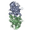
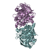
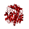
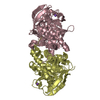
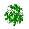
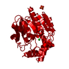
| Title | Intracellular subtilisin precursor from B. clausii | ||||||
 Components Components | (INTRACELLULAR SUBTILISIN ...) x 2 | ||||||
 Keywords Keywords | HYDROLASE / SERINE PROTEASE / INTRACELLULAR PROTEINASE REGULATION | ||||||
| Function / homology |  Function and homology information Function and homology informationserine-type endopeptidase activity / proteolysis / metal ion binding / identical protein binding Similarity search - Function | ||||||
| Biological species |  BACILLUS CLAUSII (bacteria) BACILLUS CLAUSII (bacteria) | ||||||
| Method |  X-RAY DIFFRACTION / SYNCHROTRON / MOLECULAR REPLACEMENT / Resolution: 1.56 Å X-RAY DIFFRACTION / SYNCHROTRON / MOLECULAR REPLACEMENT / Resolution: 1.56 Å | ||||||
 Authors Authors | Vedodova, J. / Gamble, M. / Ariza, A. / Dodson, E. / Jones, D.D. / Wilson, K.S. | ||||||
 Citation Citation | Journal: Structure / Year: 2010 Title: Crystal structure of an intracellular subtilisin reveals novel structural features unique to this subtilisin family. Authors: Vevodova, J. / Gamble, M. / Kunze, G. / Ariza, A. / Dodson, E. / Jones, D.D. / Wilson, K.S. | ||||||
| History |
|
- Structure visualization
| Structure viewer | Molecule: MolmilJmol/JSmol |
|---|
- Downloads & links
-Download
| PDBx/mmCIF format | 2x8j.cif.gz | 388.3 KB | Display | PDBx/mmCIF format |
|---|---|---|---|---|
| PDB format | pdb2x8j.ent.gz | 315.3 KB | Display | PDB format |
| PDBx/mmJSON format | 2x8j.json.gz | Tree view | PDBx/mmJSON format | |
| Others |  Other downloads Other downloads |
-Validation report
| Arichive directory | https://data.pdbj.org/pub/pdb/validation_reports/x8/2x8jftp://data.pdbj.org/pub/pdb/validation_reports/x8/2x8j | HTTPS FTP |
|---|
-Related structure data
| Related structure data |  2wv7C  2wwtSC C: citing same article ( S: Starting model for refinement |
|---|---|
| Similar structure data |


-Links
 PDBj
PDBj

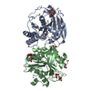
- Assembly
| Deposited unit | 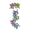
| ||||||||||||
|---|---|---|---|---|---|---|---|---|---|---|---|---|---|
| 1 | 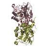
| ||||||||||||
| 2 | 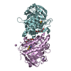
| ||||||||||||
| 3 | 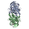
| ||||||||||||
| Unit cell |
| ||||||||||||
| Noncrystallographic symmetry (NCS) | NCS oper:
|
-Components
-INTRACELLULAR SUBTILISIN ... , 2 types, 6 molecules ACDEFB
| #1: Protein | Mass: 34717.746 Da / Num. of mol.: 5 / Mutation: YES Source method: isolated from a genetically manipulated source Details: THESE CHAINS CONTAIN 1 OXIDISED CYS / Source: (gene. exp.) BACILLUS CLAUSII (bacteria)Description: ISOLATED FROM A NOVOZYMES STRAIN B. CLAUSII STRAIN -STRAIN NUMBER AVAILABLE ON REQUEST. Plasmid: PET22B / Production host: #2: Protein | | Mass: 34733.746 Da / Num. of mol.: 1 / Mutation: YES Source method: isolated from a genetically manipulated source Details: THIS CHAIN CONTAINS 2 OXIDISED CYS / Source: (gene. exp.) BACILLUS CLAUSII (bacteria)Description: ISOLATED FROM A NOVOZYMES STRAIN B. CLAUSII STRAIN -STRAIN NUMBER AVAILABLE ON REQUEST. Plasmid: PET22B / Production host: |
|---|
-Non-polymers , 4 types, 1515 molecules 


| #3: Chemical | ChemComp-NA /  Mass: 22.990 Da / Num. of mol.: 6 / Source method: obtained synthetically / Formula: Na Mass: 22.990 Da / Num. of mol.: 6 / Source method: obtained synthetically / Formula: Na#4: Chemical | ChemComp-1PE /  Mass: 238.278 Da / Num. of mol.: 6 / Source method: obtained synthetically / Formula: C10H22O6 / Comment: precipitant*YM Mass: 238.278 Da / Num. of mol.: 6 / Source method: obtained synthetically / Formula: C10H22O6 / Comment: precipitant*YM#5: Chemical |  Mass: 92.094 Da / Num. of mol.: 2 / Source method: obtained synthetically / Formula: C3H8O3 Mass: 92.094 Da / Num. of mol.: 2 / Source method: obtained synthetically / Formula: C3H8O3#6: Water | ChemComp-HOH / | Mass: 18.015 Da / Num. of mol.: 1501 / Source method: isolated from a natural source / Formula: H2O |
|---|
-Details
| Compound details | ENGINEERED RESIDUE IN CHAIN A, SER 250 TO ALA ENGINEERED RESIDUE IN CHAIN B, SER 250 TO ALA ...ENGINEERED |
|---|
-Experimental details
-Experiment
| Experiment | Method: X-RAY DIFFRACTION / Number of used crystals: 1 |
|---|
- Sample preparation
| Crystal | Density Matthews: 2.49 Å3/Da / Density % sol: 48.5 % / Description: BASED ON PREVIOUSLY DETERMINED STRUCTURE |
|---|---|
| Crystal grow | Method: vapor diffusion, hanging drop / pH: 5.9 Details: PEG 4K GLYCEROL 0.1M SODIUM CACODYLATE PH 5.9 0.2M SODIUM ACETATE. |
-Data collection
| Diffraction | Mean temperature: 100 K |
|---|---|
| Diffraction source | Source: SYNCHROTRON / Site: Diamond  / Beamline: I03 / Wavelength: 0.9805 / Beamline: I03 / Wavelength: 0.9805 |
| Detector | Type: ADSC CCD / Detector: CCD / Date: Jan 25, 2010 |
| Radiation | Protocol: SINGLE WAVELENGTH / Monochromatic (M) / Laue (L): M / Scattering type: x-ray |
| Radiation wavelength | Wavelength: 0.9805 Å / Relative weight: 1 |
| Reflection | Resolution: 1.56→108.4 Å / Num. obs: 269002 / % possible obs: 100 % / Observed criterion σ(I): 0 / Redundancy: 7.1 % / Biso Wilson estimate: 21.9 Å2 / Rmerge(I) obs: 0.077 / Net I/σ(I): 13.4 |
| Reflection shell | Resolution: 1.56→1.64 Å / Redundancy: 5.5 % / Rmerge(I) obs: 1.08 / Mean I/σ(I) obs: 1.6 / % possible all: 100 |
- Processing
| Software |
| ||||||||||||||||||||||||||||||||||||||||||||||||||||||||||||||||||||||||||||||||||||||||||||||||||||||||||||||||||||||||||||||||||||||||||||||||||||||||||||||||||||||||||||||||||||||
|---|---|---|---|---|---|---|---|---|---|---|---|---|---|---|---|---|---|---|---|---|---|---|---|---|---|---|---|---|---|---|---|---|---|---|---|---|---|---|---|---|---|---|---|---|---|---|---|---|---|---|---|---|---|---|---|---|---|---|---|---|---|---|---|---|---|---|---|---|---|---|---|---|---|---|---|---|---|---|---|---|---|---|---|---|---|---|---|---|---|---|---|---|---|---|---|---|---|---|---|---|---|---|---|---|---|---|---|---|---|---|---|---|---|---|---|---|---|---|---|---|---|---|---|---|---|---|---|---|---|---|---|---|---|---|---|---|---|---|---|---|---|---|---|---|---|---|---|---|---|---|---|---|---|---|---|---|---|---|---|---|---|---|---|---|---|---|---|---|---|---|---|---|---|---|---|---|---|---|---|---|---|---|---|
| Refinement | Method to determine structure: MOLECULAR REPLACEMENT Starting model: PDB ENTRY 2WWT Resolution: 1.56→75.98 Å / Cor.coef. Fo:Fc: 0.968 / Cor.coef. Fo:Fc free: 0.952 / SU B: 1.958 / SU ML: 0.07 / Cross valid method: THROUGHOUT / ESU R: 0.085 / ESU R Free: 0.09 / Stereochemistry target values: MAXIMUM LIKELIHOOD / Details: HYDROGENS HAVE BEEN ADDED IN THE RIDING POSITIONS.
| ||||||||||||||||||||||||||||||||||||||||||||||||||||||||||||||||||||||||||||||||||||||||||||||||||||||||||||||||||||||||||||||||||||||||||||||||||||||||||||||||||||||||||||||||||||||
| Solvent computation | Ion probe radii: 0.8 Å / Shrinkage radii: 0.8 Å / VDW probe radii: 1.4 Å / Solvent model: MASK | ||||||||||||||||||||||||||||||||||||||||||||||||||||||||||||||||||||||||||||||||||||||||||||||||||||||||||||||||||||||||||||||||||||||||||||||||||||||||||||||||||||||||||||||||||||||
| Displacement parameters | Biso mean: 19.843 Å2
| ||||||||||||||||||||||||||||||||||||||||||||||||||||||||||||||||||||||||||||||||||||||||||||||||||||||||||||||||||||||||||||||||||||||||||||||||||||||||||||||||||||||||||||||||||||||
| Refinement step | Cycle: LAST / Resolution: 1.56→75.98 Å
| ||||||||||||||||||||||||||||||||||||||||||||||||||||||||||||||||||||||||||||||||||||||||||||||||||||||||||||||||||||||||||||||||||||||||||||||||||||||||||||||||||||||||||||||||||||||
| Refine LS restraints |
|