 Movie
Movie Controller

[English] 日本語
 Yorodumi
- PDB-6sqs: Crystal structure of cat phospho-Ser429 MDM2 RING domain bound to... -
+ Open data

- Basic information
| Entry | Database: PDB / ID: 6sqs | |||||||||
|---|---|---|---|---|---|---|---|---|---|---|
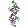
| Title | Crystal structure of cat phospho-Ser429 MDM2 RING domain bound to UbcH5B-Ub | |||||||||
 Components Components |
| |||||||||
 Keywords Keywords | LIGASE / MDM2 / MDMX / E3 / E2 / ubiquitin ligase / ubiquitin / phosphorylation | |||||||||
| Function / homology |  Function and homology information Function and homology informationregulation of biological quality / (E3-independent) E2 ubiquitin-conjugating enzyme / Formation of the ternary complex, and subsequently, the 43S complex / E2 ubiquitin-conjugating enzyme / Ribosomal scanning and start codon recognition / Translation initiation complex formation / ubiquitin conjugating enzyme activity / SARS-CoV-1 modulates host translation machinery / Peptide chain elongation / Selenocysteine synthesis ...regulation of biological quality / (E3-independent) E2 ubiquitin-conjugating enzyme / Formation of the ternary complex, and subsequently, the 43S complex / E2 ubiquitin-conjugating enzyme / Ribosomal scanning and start codon recognition / Translation initiation complex formation / ubiquitin conjugating enzyme activity / SARS-CoV-1 modulates host translation machinery / Peptide chain elongation / Selenocysteine synthesis / Formation of a pool of free 40S subunits / Eukaryotic Translation Termination / Response of EIF2AK4 (GCN2) to amino acid deficiency / SRP-dependent cotranslational protein targeting to membrane / Viral mRNA Translation / Maturation of protein E / Maturation of protein E / Nonsense Mediated Decay (NMD) independent of the Exon Junction Complex (EJC) / GTP hydrolysis and joining of the 60S ribosomal subunit / ER Quality Control Compartment (ERQC) / Myoclonic epilepsy of Lafora / FLT3 signaling by CBL mutants / Prevention of phagosomal-lysosomal fusion / L13a-mediated translational silencing of Ceruloplasmin expression / IRAK2 mediated activation of TAK1 complex / Alpha-protein kinase 1 signaling pathway / Glycogen synthesis / IRAK1 recruits IKK complex / IRAK1 recruits IKK complex upon TLR7/8 or 9 stimulation / Membrane binding and targetting of GAG proteins / Endosomal Sorting Complex Required For Transport (ESCRT) / Regulation of TBK1, IKKε (IKBKE)-mediated activation of IRF3, IRF7 / Negative regulation of FLT3 / PTK6 Regulates RTKs and Their Effectors AKT1 and DOK1 / Regulation of TBK1, IKKε-mediated activation of IRF3, IRF7 upon TLR3 ligation / Constitutive Signaling by NOTCH1 HD Domain Mutants / IRAK2 mediated activation of TAK1 complex upon TLR7/8 or 9 stimulation / NOTCH2 Activation and Transmission of Signal to the Nucleus / Major pathway of rRNA processing in the nucleolus and cytosol / TICAM1,TRAF6-dependent induction of TAK1 complex / TICAM1-dependent activation of IRF3/IRF7 / APC/C:Cdc20 mediated degradation of Cyclin B / ribonucleoprotein complex binding / Regulation of FZD by ubiquitination / Downregulation of ERBB4 signaling / p75NTR recruits signalling complexes / protein K48-linked ubiquitination / APC-Cdc20 mediated degradation of Nek2A / InlA-mediated entry of Listeria monocytogenes into host cells / Regulation of pyruvate metabolism / TRAF6-mediated induction of TAK1 complex within TLR4 complex / TRAF6 mediated IRF7 activation in TLR7/8 or 9 signaling / Regulation of innate immune responses to cytosolic DNA / NF-kB is activated and signals survival / Nonsense Mediated Decay (NMD) enhanced by the Exon Junction Complex (EJC) / Downregulation of ERBB2:ERBB3 signaling / NRIF signals cell death from the nucleus / Pexophagy / VLDLR internalisation and degradation / protein autoubiquitination / Regulation of PTEN localization / Activated NOTCH1 Transmits Signal to the Nucleus / Regulation of BACH1 activity / MAP3K8 (TPL2)-dependent MAPK1/3 activation / Translesion synthesis by REV1 / Synthesis of active ubiquitin: roles of E1 and E2 enzymes / InlB-mediated entry of Listeria monocytogenes into host cell / Translesion synthesis by POLK / Activation of IRF3, IRF7 mediated by TBK1, IKKε (IKBKE) / Downregulation of TGF-beta receptor signaling / Josephin domain DUBs / TICAM1, RIP1-mediated IKK complex recruitment / JNK (c-Jun kinases) phosphorylation and activation mediated by activated human TAK1 / Translesion synthesis by POLI / Gap-filling DNA repair synthesis and ligation in GG-NER / IKK complex recruitment mediated by RIP1 / Regulation of activated PAK-2p34 by proteasome mediated degradation / TGF-beta receptor signaling in EMT (epithelial to mesenchymal transition) / TNFR1-induced NF-kappa-B signaling pathway / PINK1-PRKN Mediated Mitophagy / TCF dependent signaling in response to WNT / cytosolic ribosome / Autodegradation of Cdh1 by Cdh1:APC/C / N-glycan trimming in the ER and Calnexin/Calreticulin cycle / APC/C:Cdc20 mediated degradation of Securin / activated TAK1 mediates p38 MAPK activation / Regulation of NF-kappa B signaling / positive regulation of mitotic cell cycle / Asymmetric localization of PCP proteins / Ubiquitin-dependent degradation of Cyclin D / Regulation of signaling by CBL / NIK-->noncanonical NF-kB signaling / NOTCH3 Activation and Transmission of Signal to the Nucleus / SCF-beta-TrCP mediated degradation of Emi1 / Negative regulators of DDX58/IFIH1 signaling / Deactivation of the beta-catenin transactivating complex / TNFR2 non-canonical NF-kB pathway / Negative regulation of FGFR3 signaling / AUF1 (hnRNP D0) binds and destabilizes mRNA / Fanconi Anemia Pathway Similarity search - Function | |||||||||
| Biological species |   Homo sapiens (human) Homo sapiens (human) | |||||||||
| Method |  X-RAY DIFFRACTION / SYNCHROTRON / MOLECULAR REPLACEMENT / Resolution: 1.83 Å X-RAY DIFFRACTION / SYNCHROTRON / MOLECULAR REPLACEMENT / Resolution: 1.83 Å | |||||||||
 Authors Authors | Magnussen, H.M. / Ahmed, S.F. / Huang, D.T. | |||||||||
| Funding support |  United Kingdom, United Kingdom,  Belgium, 2items Belgium, 2items
| |||||||||
 Citation Citation | Journal: Nat Commun / Year: 2020 Title: Structural basis for DNA damage-induced phosphoregulation of MDM2 RING domain. Authors: Magnussen, H.M. / Ahmed, S.F. / Sibbet, G.J. / Hristova, V.A. / Nomura, K. / Hock, A.K. / Archibald, L.J. / Jamieson, A.G. / Fushman, D. / Vousden, K.H. / Weissman, A.M. / Huang, D.T. | |||||||||
| History |
|
- Structure visualization
| Structure viewer | Molecule: MolmilJmol/JSmol |
|---|
- Downloads & links
-Download
| PDBx/mmCIF format | 6sqs.cif.gz | 130.1 KB | Display | PDBx/mmCIF format |
|---|---|---|---|---|
| PDB format | pdb6sqs.ent.gz | 99.4 KB | Display | PDB format |
| PDBx/mmJSON format | 6sqs.json.gz | Tree view | PDBx/mmJSON format | |
| Others |  Other downloads Other downloads |
-Validation report
| Summary document | 6sqs_validation.pdf.gz | 421.4 KB | Display | wwPDB validaton report |
|---|---|---|---|---|
| Full document | 6sqs_full_validation.pdf.gz | 421.6 KB | Display | |
| Data in XML | 6sqs_validation.xml.gz | 13 KB | Display | |
| Data in CIF | 6sqs_validation.cif.gz | 19.7 KB | Display | |
| Arichive directory | https://data.pdbj.org/pub/pdb/validation_reports/sq/6sqsftp://data.pdbj.org/pub/pdb/validation_reports/sq/6sqs | HTTPS FTP |
-Related structure data
| Related structure data |  6sqoC  6sqpC  6sqrC  5mnjS C: citing same article ( S: Starting model for refinement |
|---|---|
| Similar structure data |


-Links
 PDBj
PDBj


- Assembly
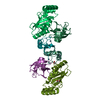
| Deposited unit | 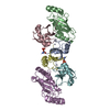
| ||||||||
|---|---|---|---|---|---|---|---|---|---|
| 1 |
| ||||||||
| Unit cell |
|
-Components
| #1: Protein | Mass: 7287.754 Da / Num. of mol.: 2 / Mutation: G443T Source method: isolated from a genetically manipulated source Details: Residues 422-491 and contains G443T mutation. S429 is phosphorylated. GS at the N-terminus resulted from cloning. Source: (gene. exp.)  References: UniProt: Q7YRZ8, RING-type E3 ubiquitin transferase #2: Protein | Mass: 16720.186 Da / Num. of mol.: 2 / Mutation: S22R, C85K Source method: isolated from a genetically manipulated source Details: Contains S22R and C85K mutations. K85 in Chains B and E form a covalent bond with G76 in Chains C and F, respectively. Source: (gene. exp.) Homo sapiens (human) / Gene: UBE2D2, PUBC1, UBC4, UBC5B, UBCH4, UBCH5B / Production host: References: UniProt: P62837, E2 ubiquitin-conjugating enzyme, (E3-independent) E2 ubiquitin-conjugating enzyme #3: Protein | Mass: 8663.908 Da / Num. of mol.: 2 Source method: isolated from a genetically manipulated source Details: Contains a GSGGS at the N-terminus resulted from cloning. G76 in Chains C and F form a covalent bond with K85 sidechain in Chains B and E, respectively. Source: (gene. exp.) Homo sapiens (human) / Gene: RPS27A / Production host: #4: Chemical | ChemComp-ZN /   Mass: 65.409 Da / Num. of mol.: 4 / Source method: obtained synthetically / Formula: Zn Mass: 65.409 Da / Num. of mol.: 4 / Source method: obtained synthetically / Formula: Zn#5: Water | ChemComp-HOH / |  Mass: 18.015 Da / Num. of mol.: 180 / Source method: isolated from a natural source / Formula: H2O Mass: 18.015 Da / Num. of mol.: 180 / Source method: isolated from a natural source / Formula: H2OHas ligand of interest | Y | Has protein modification | Y | |
|---|
-Experimental details
-Experiment
| Experiment | Method: X-RAY DIFFRACTION / Number of used crystals: 1 |
|---|
- Sample preparation
| Crystal | Density Matthews: 2.43 Å3/Da / Density % sol: 49.43 % |
|---|---|
| Crystal grow | Temperature: 292 K / Method: vapor diffusion Details: 0.1 M Tris-HCl, pH 8.0, 15% (w/v) PEG 2000 MME and 0.1 M KCl |
-Data collection
| Diffraction | Mean temperature: 100 K / Serial crystal experiment: N |
|---|---|
| Diffraction source | Source: SYNCHROTRON / Site: Diamond / Beamline: I04-1 / Wavelength: 0.916 Å |
| Detector | Type: DECTRIS PILATUS 6M-F / Detector: PIXEL / Date: Nov 25, 2018 |
| Radiation | Monochromator: 0.916 / Protocol: SINGLE WAVELENGTH / Monochromatic (M) / Laue (L): M / Scattering type: x-ray |
| Radiation wavelength | Wavelength: 0.916 Å / Relative weight: 1 |
| Reflection | Resolution: 1.83→29.2 Å / Num. obs: 49057 / % possible obs: 95.8 % / Redundancy: 1.8 % / CC1/2: 0.996 / Net I/σ(I): 10.2 |
| Reflection shell | Resolution: 1.83→1.88 Å / Num. unique obs: 3293 / CC1/2: 0.539 |
- Processing
| Software |
| ||||||||||||||||
|---|---|---|---|---|---|---|---|---|---|---|---|---|---|---|---|---|---|
| Refinement | Method to determine structure: MOLECULAR REPLACEMENT Starting model: 5MNJ Resolution: 1.83→29.2 Å / Cross valid method: FREE R-VALUE
| ||||||||||||||||
| Refinement step | Cycle: LAST / Resolution: 1.83→29.2 Å
|
