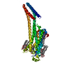
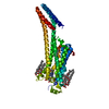
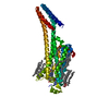
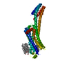
[English] 日本語
Yorodumi
- PDB-5k2c: 1.9 angstrom A2a adenosine receptor structure with sulfur SAD pha... -

+
Open data


ID or keywords:

Loading...

-
Basic information

Entry
Database: PDB / ID: 5k2c
Title1.9 angstrom A2a adenosine receptor structure with sulfur SAD phasing and phase extension using XFEL data
ComponentsAdenosine receptor A2a/Soluble cytochrome b562 chimera
KeywordsMEMBRANE PROTEIN / A2a Adenosine Receptor / sulfur SAD / GPCR / Structural Genomics / PSI-Biology / GPCR Network
Function / homology
Function and homology information


positive regulation of acetylcholine secretion, neurotransmission / regulation of norepinephrine secretion / positive regulation of circadian sleep/wake cycle, sleep / Adenosine P1 receptors / negative regulation of alpha-beta T cell activation / G protein-coupled adenosine receptor activity / G protein-coupled adenosine receptor signaling pathway / response to purine-containing compound / sensory perception / positive regulation of urine volume ...positive regulation of acetylcholine secretion, neurotransmission / regulation of norepinephrine secretion / positive regulation of circadian sleep/wake cycle, sleep / Adenosine P1 receptors / negative regulation of alpha-beta T cell activation / G protein-coupled adenosine receptor activity / G protein-coupled adenosine receptor signaling pathway / response to purine-containing compound / sensory perception / positive regulation of urine volume / NGF-independant TRKA activation / Surfactant metabolism / protein kinase C-activating G protein-coupled receptor signaling pathway / synaptic transmission, dopaminergic / synaptic transmission, cholinergic / inhibitory postsynaptic potential / negative regulation of vascular permeability / type 5 metabotropic glutamate receptor binding / positive regulation of glutamate secretion / blood circulation / intermediate filament / response to caffeine / eating behavior / presynaptic active zone / alpha-actinin binding / membrane depolarization / regulation of calcium ion transport / asymmetric synapse / axolemma / response to inorganic substance / cellular defense response / prepulse inhibition / phagocytosis / positive regulation of apoptotic signaling pathway / response to amphetamine / excitatory postsynaptic potential / presynaptic modulation of chemical synaptic transmission / positive regulation of synaptic transmission, glutamatergic / neuron projection morphogenesis / regulation of mitochondrial membrane potential / locomotory behavior / synaptic transmission, glutamatergic / central nervous system development / positive regulation of long-term synaptic potentiation / astrocyte activation / apoptotic signaling pathway / positive regulation of synaptic transmission, GABAergic / positive regulation of protein secretion / adenylate cyclase-modulating G protein-coupled receptor signaling pathway / adenylate cyclase-activating G protein-coupled receptor signaling pathway / negative regulation of inflammatory response / vasodilation / blood coagulation / cell-cell signaling / presynaptic membrane / G alpha (s) signalling events / postsynaptic membrane / negative regulation of neuron apoptotic process / electron transfer activity / periplasmic space / calmodulin binding / response to xenobiotic stimulus / inflammatory response / iron ion binding / negative regulation of cell population proliferation / dendrite / neuronal cell body / lipid binding / glutamatergic synapse / apoptotic process / heme binding / protein-containing complex binding / regulation of DNA-templated transcription / enzyme binding / membrane / identical protein binding / plasma membrane
Similarity search - Function
Adenosine A2A receptor / Adenosine receptor / Cytochrome b562 / Cytochrome b562 / Cytochrome c/b562 / Serpentine type 7TM GPCR chemoreceptor Srsx / G-protein coupled receptors family 1 signature. / G protein-coupled receptor, rhodopsin-like / GPCR, rhodopsin-like, 7TM / G-protein coupled receptors family 1 profile. / 7 transmembrane receptor (rhodopsin family)
Similarity search - Domain/homology
CHOLESTEROL / OLEIC ACID / (2R)-2,3-dihydroxypropyl (9Z)-octadec-9-enoate / DI(HYDROXYETHYL)ETHER / Chem-ZMA / Soluble cytochrome b562 / Adenosine receptor A2a
Similarity search - Component
Biological speciesHomo sapiens (human)
Escherichia coli (E. coli)
MethodX-RAY DIFFRACTION / FREE ELECTRON LASER / SAD / Resolution: 1.9 Å
AuthorsBatyuk, A. / Galli, L. / Ishchenko, A. / Han, G.W. / Gati, C. / Popov, P. / Lee, M.-Y. / Stauch, B. / White, T.A. / Barty, A. ...Batyuk, A. / Galli, L. / Ishchenko, A. / Han, G.W. / Gati, C. / Popov, P. / Lee, M.-Y. / Stauch, B. / White, T.A. / Barty, A. / Aquila, A. / Hunter, M.S. / Liang, M. / Boutet, S. / Pu, M. / Liu, Z.-J. / Nelson, G. / James, D. / Li, C. / Zhao, Y. / Spence, J.C.H. / Liu, W. / Fromme, P. / Katritch, V. / Weierstall, U. / Stevens, R.C. / Cherezov, V. / GPCR Network (GPCR)
CitationJournal: Sci Adv / Year: 2016
Title: Native phasing of x-ray free-electron laser data for a G protein-coupled receptor.
Authors: Batyuk, A. / Galli, L. / Ishchenko, A. / Han, G.W. / Gati, C. / Popov, P.A. / Lee, M.Y. / Stauch, B. / White, T.A. / Barty, A. / Aquila, A. / Hunter, M.S. / Liang, M. / Boutet, S. / Pu, M. / ...Authors: Batyuk, A. / Galli, L. / Ishchenko, A. / Han, G.W. / Gati, C. / Popov, P.A. / Lee, M.Y. / Stauch, B. / White, T.A. / Barty, A. / Aquila, A. / Hunter, M.S. / Liang, M. / Boutet, S. / Pu, M. / Liu, Z.J. / Nelson, G. / James, D. / Li, C. / Zhao, Y. / Spence, J.C. / Liu, W. / Fromme, P. / Katritch, V. / Weierstall, U. / Stevens, R.C. / Cherezov, V.
History
DepositionMay 18, 2016Deposition site: RCSB / Processing site: RCSB
Revision 1.0Sep 21, 2016Provider: repository / Type: Initial release
Revision 1.1Oct 12, 2016Group: Database references
Revision 1.2Nov 23, 2016Group: Structure summary
Revision 1.3Nov 22, 2017Group: Refinement description / Category: software
Revision 1.4Feb 14, 2018Group: Data collection / Category: diffrn_source
Item: _diffrn_source.pdbx_synchrotron_beamline / _diffrn_source.pdbx_synchrotron_site
Revision 1.5Nov 28, 2018Group: Data collection
Category: diffrn / pdbx_serial_crystallography_data_reduction ...diffrn / pdbx_serial_crystallography_data_reduction / pdbx_serial_crystallography_measurement / pdbx_serial_crystallography_sample_delivery / pdbx_serial_crystallography_sample_delivery_injection
Item: _diffrn.pdbx_serial_crystal_experiment

-
Structure visualization

Structure viewerMolecule:
MolmilJmol/JSmol

Downloads & links

-
Assembly

Deposited unit
A: Adenosine receptor A2a/Soluble cytochrome b562 chimera
hetero molecules


Theoretical massNumber of molelcules
Total (without water)58,27328
Polymers49,9741
Non-polymers8,29927
Water1,892105
1


  • Idetical with deposited unit
  • defined by author
TypeNameSymmetry operationNumber
identity operation1_555x,y,z1
Unit cell
Length a, b, c (Å)40.360, 180.740, 142.800
Angle α, β, γ (deg.)90.00, 90.00, 90.00
Int Tables number20
Space group name H-MC2221

-
Components

-
Protein , 1 types, 1 molecules A

#1: Protein Adenosine receptor A2a/Soluble cytochrome b562 chimera / Cytochrome b-562


Mass: 49974.281 Da / Num. of mol.: 1
Source method: isolated from a genetically manipulated source
Source: (gene. exp.) Homo sapiens, Escherichia coli / Gene: ADORA2A, ADORA2, cybC / Production host: 
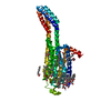
Spodoptera frugiperda (fall armyworm) / References: UniProt: P29274, UniProt: P0ABE7

-
Non-polymers , 7 types, 132 molecules

#2: Chemical ChemComp-ZMA / 4-{2-[(7-amino-2-furan-2-yl[1,2,4]triazolo[1,5-a][1,3,5]triazin-5-yl)amino]ethyl}phenol / ZM-241,385


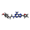
Mass: 337.336 Da / Num. of mol.: 1 / Source method: obtained synthetically / Formula: C16H15N7O2 / Comment: antagonist*YM
#3: Chemical ChemComp-NA / SODIUM ION


Mass: 22.990 Da / Num. of mol.: 1 / Source method: obtained synthetically / Formula: Na
#4: Chemical ChemComp-CLR / CHOLESTEROL / Cholesterol


Mass: 386.654 Da / Num. of mol.: 3 / Source method: obtained synthetically / Formula: C27H46O
#5: Chemical
ChemComp-OLC / (2R)-2,3-dihydroxypropyl (9Z)-octadec-9-enoate / 1-Oleoyl-R-glycerol


Mass: 356.540 Da / Num. of mol.: 10 / Source method: obtained synthetically / Formula: C21H40O4
#6: Chemical
ChemComp-OLA / OLEIC ACID / Oleic acid


Mass: 282.461 Da / Num. of mol.: 11 / Source method: obtained synthetically / Formula: C18H34O2
#7: Chemical ChemComp-PEG / DI(HYDROXYETHYL)ETHER / Diethylene glycol


Mass: 106.120 Da / Num. of mol.: 1 / Source method: obtained synthetically / Formula: C4H10O3
#8: Water ChemComp-HOH / water / Water


Mass: 18.015 Da / Num. of mol.: 105 / Source method: isolated from a natural source / Formula: H2O

-
Experimental details

-
Experiment

ExperimentMethod: X-RAY DIFFRACTION / Number of used crystals: 1

-
Sample preparation

CrystalDensity Matthews: 2.61 Å3/Da / Density % sol: 52.79 %
Crystal growTemperature: 293 K / Method: lipidic cubic phase / pH: 5
Details: 28 % (v/v) PEG 400, 40 mM sodium thiocyanate and 100 mM sodium citrate buffer pH 5.0

-
Data collection

DiffractionMean temperature: 293 K / Serial crystal experiment: Y
Diffraction sourceSource: FREE ELECTRON LASER / Site: SLAC LCLS / Beamline: CXI / Wavelength: 1.27 Å
DetectorType: CS-PAD CXI-1 / Detector: PIXEL / Date: Apr 1, 2015 / Details: K-B mirrors
RadiationProtocol: SINGLE WAVELENGTH / Monochromatic (M) / Laue (L): M / Scattering type: x-ray
Radiation wavelengthWavelength: 1.27 Å / Relative weight: 1
ReflectionResolution: 1.9→24 Å / Num. obs: 41882 / % possible obs: 100 % / Redundancy: 291 % / Biso Wilson estimate: 46.9 Å2 / Net I/σ(I): 6
Reflection shellResolution: 1.9→2 Å / Redundancy: 62 % / Mean I/σ(I) obs: 0.6 / % possible all: 100
Serial crystallography measurementFocal spot size: 1.5 µm2 / Photons per pulse: 64000000000 Tphotons/pulse / Pulse duration: 35 fsec. / Pulse energy: 2.18 µJ / Pulse photon energy: 9.8 keV / XFEL pulse repetition rate: 120 Hz
Serial crystallography sample deliveryMethod: injection
Serial crystallography sample delivery injectionFlow rate: 220 µL/min
Serial crystallography data reductionCrystal hits: 232283 / Frames indexed: 72735 / Frames total: 948961

-
Processing

Software
NameVersionClassification
PHENIX1.10_2155refinement
CrystFELdata reduction
CrystFELdata scaling
SHELXDphasing
PHASERphasing
RefinementMethod to determine structure: SAD / Resolution: 1.9→23.437 Å / SU ML: 0.31 / Cross valid method: THROUGHOUT / σ(F): 1.33 / Phase error: 22.75 / Stereochemistry target values: MLHL
RfactorNum. reflection% reflection
Rfree0.2079 1988 4.75 %
Rwork0.1726 --
obs0.1743 41828 99.95 %
Solvent computationShrinkage radii: 0.9 Å / VDW probe radii: 1.11 Å / Solvent model: FLAT BULK SOLVENT MODEL
Refinement stepCycle: LAST / Resolution: 1.9→23.437 Å
ProteinNucleic acidLigandSolventTotal
Num. atoms3013 0 415 105 3533
Refine LS restraints
Refine-IDTypeDev idealNumber
X-RAY DIFFRACTIONf_bond_d0.013598
X-RAY DIFFRACTIONf_angle_d1.1764848
X-RAY DIFFRACTIONf_dihedral_angle_d15.0252166
X-RAY DIFFRACTIONf_chiral_restr0.059558
X-RAY DIFFRACTIONf_plane_restr0.006576
LS refinement shell
Resolution (Å)Rfactor RfreeNum. reflection RfreeRfactor RworkNum. reflection RworkRefine-ID% reflection obs (%)
1.9-1.94750.43351180.45192815X-RAY DIFFRACTION100
1.9475-2.00010.41541710.4182794X-RAY DIFFRACTION100
2.0001-2.05890.33051260.31672797X-RAY DIFFRACTION100
2.0589-2.12540.28881350.24982835X-RAY DIFFRACTION100
2.1254-2.20130.22181370.22392797X-RAY DIFFRACTION100
2.2013-2.28930.23511580.19662795X-RAY DIFFRACTION100
2.2893-2.39340.22731240.17222860X-RAY DIFFRACTION100
2.3934-2.51950.19541350.15242828X-RAY DIFFRACTION100
2.5195-2.67710.17951670.14162807X-RAY DIFFRACTION100
2.6771-2.88350.151350.13632861X-RAY DIFFRACTION100
2.8835-3.1730.21581440.13992826X-RAY DIFFRACTION100
3.173-3.63070.1651460.14392882X-RAY DIFFRACTION100
3.6307-4.56880.18781350.14422920X-RAY DIFFRACTION100
4.5688-23.43860.21881570.18643023X-RAY DIFFRACTION100
Refinement TLS params.

Method: refined / Refine-ID: X-RAY DIFFRACTION

IDL112)L122)L132)L222)L232)L332)S11 (Å °)S12 (Å °)S13 (Å °)S21 (Å °)S22 (Å °)S23 (Å °)S31 (Å °)S32 (Å °)S33 (Å °)T112)T122)T132)T222)T232)T332)Origin x (Å)Origin y (Å)Origin z (Å)
11.3440.25440.14452.197-0.14631.7016-0.00820.0495-0.0212-0.07130.0127-0.05490.05220.0898-0.00530.2298-0.00890.01520.2809-0.01070.2133.902184.328850.2567
23.6718-2.86530.99457.97563.03152.91040.3390.3484-0.26070.45110.15460.20840.2985-0.0893-0.56710.7951-0.1141-0.07580.55610.10811.2393-20.770935.76851.4366
31.2956-0.8150.32925.3643-0.49181.73080.0061-0.1086-0.15120.44160.01650.13080.2480.0098-0.0140.2576-0.0310.04560.2658-0.01080.1989-1.637479.219860.4654
Refinement TLS group
IDRefine-IDRefine TLS-IDSelection details
1X-RAY DIFFRACTION1chain 'A' and (resid -2 through 208 )
2X-RAY DIFFRACTION2chain 'A' and (resid 1001 through 1106 )
3X-RAY DIFFRACTION3chain 'A' and (resid 219 through 308)

+
About Yorodumi

-
News

-
Feb 9, 2022. New format data for meta-information of EMDB entries

New format data for meta-information of EMDB entries

  • Version 3 of the EMDB header file is now the official format.
  • The previous official version 1.9 will be removed from the archive.

Related info.:EMDB header

External links:wwPDB to switch to version 3 of the EMDB data model

-
Aug 12, 2020. Covid-19 info

Covid-19 info

URL: https://pdbj.org/emnavi/covid19.php

New page: Covid-19 featured information page in EM Navigator.

Related info.:Covid-19 info / Mar 5, 2020. Novel coronavirus structure data

+
Mar 5, 2020. Novel coronavirus structure data

Novel coronavirus structure data

Related info.:Yorodumi Speices / Aug 12, 2020. Covid-19 info

External links:COVID-19 featured content - PDBj / Molecule of the Month (242):Coronavirus Proteases

+
Jan 31, 2019. EMDB accession codes are about to change! (news from PDBe EMDB page)

EMDB accession codes are about to change! (news from PDBe EMDB page)

  • The allocation of 4 digits for EMDB accession codes will soon come to an end. Whilst these codes will remain in use, new EMDB accession codes will include an additional digit and will expand incrementally as the available range of codes is exhausted. The current 4-digit format prefixed with “EMD-” (i.e. EMD-XXXX) will advance to a 5-digit format (i.e. EMD-XXXXX), and so on. It is currently estimated that the 4-digit codes will be depleted around Spring 2019, at which point the 5-digit format will come into force.
  • The EM Navigator/Yorodumi systems omit the EMD- prefix.

Related info.:Q: What is EMD? / ID/Accession-code notation in Yorodumi/EM Navigator

External links:EMDB Accession Codes are Changing Soon! / Contact to PDBj

+
Jul 12, 2017. Major update of PDB

Major update of PDB

  • wwPDB released updated PDB data conforming to the new PDBx/mmCIF dictionary.
  • This is a major update changing the version number from 4 to 5, and with Remediation, in which all the entries are updated.
  • In this update, many items about electron microscopy experimental information are reorganized (e.g. em_software).
  • Now, EM Navigator and Yorodumi are based on the updated data.

External links:wwPDB Remediation / Enriched Model Files Conforming to OneDep Data Standards Now Available in the PDB FTP Archive

-
Yorodumi

Thousand views of thousand structures

  • Yorodumi is a browser for structure data from EMDB, PDB, SASBDB, etc.
  • This page is also the successor to EM Navigator detail page, and also detail information page/front-end page for Omokage search.
  • The word "yorodu" (or yorozu) is an old Japanese word meaning "ten thousand". "mi" (miru) is to see.

Related info.:EMDB / PDB / SASBDB / Comparison of 3 databanks / Yorodumi Search / Aug 31, 2016. New EM Navigator & Yorodumi / Yorodumi Papers / Jmol/JSmol / Function and homology information / Changes in new EM Navigator and Yorodumi

Read more