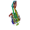
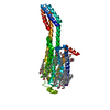
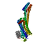
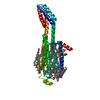
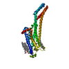
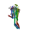
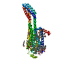
Entry Database : PDB / ID : 6aqfTitle Crystal structure of A2AAR-BRIL in complex with the antagonist ZM241385 produced from Pichia pastoris Adenosine receptor A2a,Soluble cytochrome b562,Adenosine receptor A2a Keywords / / / / / / / Function / homology Function Domain/homology Component
/ / / / / / / / / / / / / / / / / / / / / / / / / / / / / / / / / / / / / / / / / / / / / / / / / / / / / / / / / / / / / / / / / / / / / / / / / / / / / / / / / / / / / / / / / / / / / / / / / / / / / / Biological species Homo sapiens (human)Escherichia coli (E. coli)Method / / / Resolution : 2.51 Å Authors Eddy, M.T. / Lee, M.Y. / Gao, Z. / White, K. / Didenko, T. / Horst, R. / Audet, M. / Stanczak, P. / McClary, K.M. / Han, G.W. ...Eddy, M.T. / Lee, M.Y. / Gao, Z. / White, K. / Didenko, T. / Horst, R. / Audet, M. / Stanczak, P. / McClary, K.M. / Han, G.W. / Jacobson, K.A. / Stevens, R.C. / Wuthrich, K. Journal : Cell / Year : 2018Title : Allosteric Coupling of Drug Binding and Intracellular Signaling in the A2A Adenosine Receptor.Authors : Eddy, M.T. / Lee, M.Y. / Gao, Z.G. / White, K.L. / Didenko, T. / Horst, R. / Audet, M. / Stanczak, P. / McClary, K.M. / Han, G.W. / Jacobson, K.A. / Stevens, R.C. / Wuthrich, K. History Deposition Aug 19, 2017 Deposition site / Processing site Revision 1.0 Jan 10, 2018 Provider / Type Revision 1.1 Jan 17, 2018 Group / Category / citation_authorItem _citation.country / _citation.journal_abbrev ... _citation.country / _citation.journal_abbrev / _citation.journal_id_ASTM / _citation.journal_id_CSD / _citation.journal_id_ISSN / _citation.pdbx_database_id_DOI / _citation.pdbx_database_id_PubMed / _citation.title / _citation.year / _citation_author.name Revision 1.2 Jan 24, 2018 Group / Category Item _citation.journal_volume / _citation.page_first ... _citation.journal_volume / _citation.page_first / _citation.page_last / _citation.year Revision 1.3 Oct 4, 2023 Group / Database references / Refinement descriptionCategory chem_comp_atom / chem_comp_bond ... chem_comp_atom / chem_comp_bond / database_2 / pdbx_initial_refinement_model Item / _database_2.pdbx_database_accessionRevision 1.4 Oct 16, 2024 Group / Category / pdbx_modification_feature
Show all Show less
 Movie
Movie Controller

 Yorodumi
 Open data
 Basic information
 Components
 Keywords
 Function and homology information
 Homo sapiens (human)
 X-RAY DIFFRACTION /
  Authors
 Citation
 Structure visualization
 Downloads & links
 Other downloads
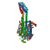

 PDBj
PDBj


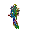
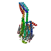
 Assembly
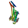
 Pichia (fungus) / References: UniProt: P29274, UniProt: P0ABE7
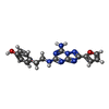


 Mass: 337.336 Da / Num. of mol.: 1 / Source method: obtained synthetically / Formula: C16H15N7O2 / Feature type: SUBJECT OF INVESTIGATION / Comment: antagonist*YM
 Mass: 386.654 Da / Num. of mol.: 3 / Source method: obtained synthetically / Formula: C27H46O
 Mass: 356.540 Da / Num. of mol.: 1 / Source method: obtained synthetically / Formula: C21H40O4 / Feature type: SUBJECT OF INVESTIGATION
 Mass: 282.461 Da / Num. of mol.: 7 / Source method: obtained synthetically / Formula: C18H34O2 / Feature type: SUBJECT OF INVESTIGATION
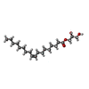
 Mass: 356.540 Da / Num. of mol.: 4 / Source method: obtained synthetically / Formula: C21H40O4 / Feature type: SUBJECT OF INVESTIGATION
 Sample preparation
 / Beamline: 23-ID-D / Wavelength: 1.033 Å
 Processing