 Movie
Movie Controller

[English] 日本語
 Yorodumi
- PDB-6so3: The interacting head motif in insect flight muscle myosin thick f... -
+ Open data

- Basic information
| Entry | Database: PDB / ID: 6so3 | ||||||
|---|---|---|---|---|---|---|---|
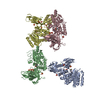
| Title | The interacting head motif in insect flight muscle myosin thick filaments | ||||||
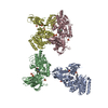
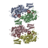
 Components Components |
| ||||||
 Keywords Keywords | MOTOR PROTEIN / myosin / thick filament / insect flight muscle | ||||||
| Biological species |  Lethocerus indicus (insect) Lethocerus indicus (insect) | ||||||
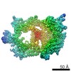
| Method | ELECTRON MICROSCOPY / helical reconstruction / cryo EM / Resolution: 6.2 Å | ||||||
 Authors Authors | Morris, E.P. / Knupp, C. / Squire, J.M. | ||||||
 Citation Citation | Journal: Biology (Basel) / Year: 2019 Title: The Interacting Head Motif Structure Does Not Explain the X-Ray Diffraction Patterns in Relaxed Vertebrate (Bony Fish) Skeletal Muscle and Insect () Flight Muscle. Authors: Carlo Knupp / Edward Morris / John M Squire /  Abstract: Unlike electron microscopy, which can achieve very high resolution but to date can only be used to study static structures, time-resolved X-ray diffraction from contracting muscles can, in principle, ...Unlike electron microscopy, which can achieve very high resolution but to date can only be used to study static structures, time-resolved X-ray diffraction from contracting muscles can, in principle, be used to follow the molecular movements involved in force generation on a millisecond timescale, albeit at moderate resolution. However, previous X-ray diffraction studies of resting muscles have come up with structures for the head arrangements in resting myosin filaments that are different from the apparently ubiquitous interacting head motif (IHM) structures found by single particle analysis of electron micrographs of isolated myosin filaments from a variety of muscle types. This head organization is supposed to represent the super-relaxed state of the myosin filaments where adenosine triphosphate (ATP) usage is minimized. Here we have tested whether the interacting head motif structures will satisfactorily explain the observed low-angle X-ray diffraction patterns from resting vertebrate (bony fish) and invertebrate (insect flight) muscles. We find that the interacting head motif does not, in fact, explain what is observed. Previous X-ray models fit the observations much better. We conclude that the X-ray diffraction evidence has been well interpreted in the past and that there is more than one ordered myosin head state in resting muscle. There is, therefore, no reason to question some of the previous X-ray diffraction results on myosin filaments; time-resolved X-ray diffraction should be a reliable way to follow crossbridge action in active muscle and may be one of the few ways to visualise the molecular changes in myosin heads on a millisecond timescale as force is actually produced. | ||||||
| History |
|
- Structure visualization
| Movie |
 Movie viewer Movie viewer |
|---|---|
| Structure viewer | Molecule: MolmilJmol/JSmol |
- Downloads & links
-Download
| PDBx/mmCIF format | 6so3.cif.gz | 822.9 KB | Display | PDBx/mmCIF format |
|---|---|---|---|---|
| PDB format | pdb6so3.ent.gz | 644.7 KB | Display | PDB format |
| PDBx/mmJSON format | 6so3.json.gz | Tree view | PDBx/mmJSON format | |
| Others |  Other downloads Other downloads |
-Validation report
| Arichive directory | https://data.pdbj.org/pub/pdb/validation_reports/so/6so3ftp://data.pdbj.org/pub/pdb/validation_reports/so/6so3 | HTTPS FTP |
|---|
-Related structure data
| Related structure data | 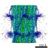 7029M M: map data used to model this data |
|---|---|
| Similar structure data |


-Links
 PDBj
PDBj
- Assembly
| Deposited unit | 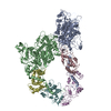
|
|---|---|
| 1 |
|
-Components
| #1: Protein | Mass: 224542.281 Da / Num. of mol.: 2 / Source method: isolated from a natural source / Source: (natural) Lethocerus indicus (insect)#2: Protein | Mass: 17628.104 Da / Num. of mol.: 2 / Source method: isolated from a natural source / Source: (natural) Lethocerus indicus (insect)#3: Protein | Mass: 21794.281 Da / Num. of mol.: 2 / Source method: isolated from a natural source / Source: (natural) Lethocerus indicus (insect) |
|---|
-Experimental details
-Experiment
| Experiment | Method: ELECTRON MICROSCOPY |
|---|---|
| EM experiment | Aggregation state: HELICAL ARRAY / 3D reconstruction method: helical reconstruction |
- Sample preparation
| Component | Name: Myosin thick filament / Type: COMPLEX / Entity ID: all / Source: NATURAL |
|---|---|
| Molecular weight | Experimental value: NO |
| Source (natural) | Organism: Lethocerus indicus (insect) |
| Buffer solution | pH: 7 |
| Specimen | Embedding applied: NO / Shadowing applied: NO / Staining applied: NO / Vitrification applied: YES |
| Vitrification | Cryogen name: ETHANE |
- Electron microscopy imaging
| Experimental equipment |  Model: Titan Krios / Image courtesy: FEI Company |
|---|---|
| Microscopy | Model: FEI TITAN KRIOS |
| Electron gun | Electron source:  FIELD EMISSION GUN / Accelerating voltage: 300 kV / Illumination mode: FLOOD BEAM FIELD EMISSION GUN / Accelerating voltage: 300 kV / Illumination mode: FLOOD BEAM |
| Electron lens | Mode: BRIGHT FIELD |
| Specimen holder | Cryogen: NITROGEN |
| Image recording | Electron dose: 65 e/Å2 / Film or detector model: DIRECT ELECTRON DE-20 (5k x 3k) |
- Processing
| CTF correction | Type: PHASE FLIPPING AND AMPLITUDE CORRECTION |
|---|---|
| Helical symmerty | Angular rotation/subunit: 33.98 ° / Axial rise/subunit: 145 Å / Axial symmetry: C4 |
| 3D reconstruction | Resolution: 6.2 Å / Resolution method: FSC 0.143 CUT-OFF / Num. of particles: 24000 / Symmetry type: HELICAL |
