 Movie
Movie Controller


+ Open data

- Basic information
| Entry | Database: PDB / ID: 6snv | ||||||
|---|---|---|---|---|---|---|---|
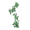
| Title | DNA mismatch repair proteins MLH1 and MLH3 | ||||||
 Components Components |
| ||||||
 Keywords Keywords | RECOMBINATION / RESOLVASE / MMR / DNA REPAIR / MEIOSIS | ||||||
| Function / homology |  Function and homology information Function and homology informationmeiotic heteroduplex formation / MutLbeta complex / MutLgamma complex / MutLalpha complex / mismatch repair complex / Mismatch repair (MMR) directed by MSH2:MSH6 (MutSalpha) / meiotic mismatch repair / mismatched DNA binding / reciprocal meiotic recombination / ATP-dependent DNA damage sensor activity ...meiotic heteroduplex formation / MutLbeta complex / MutLgamma complex / MutLalpha complex / mismatch repair complex / Mismatch repair (MMR) directed by MSH2:MSH6 (MutSalpha) / meiotic mismatch repair / mismatched DNA binding / reciprocal meiotic recombination / ATP-dependent DNA damage sensor activity / mismatch repair / ATP hydrolysis activity / mitochondrion / ATP binding / nucleus / cytoplasm Similarity search - Function | ||||||
| Biological species |  | ||||||
| Method |  X-RAY DIFFRACTION / SYNCHROTRON / MOLECULAR REPLACEMENT / Resolution: 2.5 Å X-RAY DIFFRACTION / SYNCHROTRON / MOLECULAR REPLACEMENT / Resolution: 2.5 Å | ||||||
 Authors Authors | Dai, J. / Chervy, P. / Legrand, P. / Ropars, V. / Charbonnier, J.B. | ||||||
| Funding support |  France, 1items France, 1items
| ||||||
 Citation Citation | Journal: Proc.Natl.Acad.Sci.USA / Year: 2021 Title: Molecular basis of the dual role of the Mlh1-Mlh3 endonuclease in MMR and in meiotic crossover formation. Authors: Dai, J. / Sanchez, A. / Adam, C. / Ranjha, L. / Reginato, G. / Chervy, P. / Tellier-Lebegue, C. / Andreani, J. / Guerois, R. / Ropars, V. / Le Du, M.H. / Maloisel, L. / Martini, E. / ...Authors: Dai, J. / Sanchez, A. / Adam, C. / Ranjha, L. / Reginato, G. / Chervy, P. / Tellier-Lebegue, C. / Andreani, J. / Guerois, R. / Ropars, V. / Le Du, M.H. / Maloisel, L. / Martini, E. / Legrand, P. / Thureau, A. / Cejka, P. / Borde, V. / Charbonnier, J.B. | ||||||
| History |
|
- Structure visualization
| Structure viewer | Molecule: MolmilJmol/JSmol |
|---|
- Downloads & links
-Download
| PDBx/mmCIF format | 6snv.cif.gz | 411.6 KB | Display | PDBx/mmCIF format |
|---|---|---|---|---|
| PDB format | pdb6snv.ent.gz | 338.4 KB | Display | PDB format |
| PDBx/mmJSON format | 6snv.json.gz | Tree view | PDBx/mmJSON format | |
| Others |  Other downloads Other downloads |
-Validation report
| Arichive directory | https://data.pdbj.org/pub/pdb/validation_reports/sn/6snvftp://data.pdbj.org/pub/pdb/validation_reports/sn/6snv | HTTPS FTP |
|---|
-Related structure data
| Related structure data |  6rmnC  6shxC  6snsC  4e4wS S: Starting model for refinement C: citing same article ( |
|---|---|
| Similar structure data |


-Links
 PDBj
PDBj
- Assembly
| Deposited unit | 
| ||||||||
|---|---|---|---|---|---|---|---|---|---|
| 1 | 
| ||||||||
| 2 | 
| ||||||||
| Unit cell |
|
-Components
| #1: Protein | Mass: 30764.279 Da / Num. of mol.: 2 Source method: isolated from a genetically manipulated source Source: (gene. exp.) Gene: MLH1, PMS2, YMR167W, YM8520.16 / Production host:  #2: Protein | Mass: 27767.180 Da / Num. of mol.: 2 Source method: isolated from a genetically manipulated source Source: (gene. exp.) Gene: MLH3, YPL164C, P2550 / Production host: #3: Chemical | ChemComp-ZN /   Mass: 65.409 Da / Num. of mol.: 4 / Source method: obtained synthetically / Formula: Zn / Feature type: SUBJECT OF INVESTIGATION Mass: 65.409 Da / Num. of mol.: 4 / Source method: obtained synthetically / Formula: Zn / Feature type: SUBJECT OF INVESTIGATION#4: Water | ChemComp-HOH / |  Mass: 18.015 Da / Num. of mol.: 25 / Source method: isolated from a natural source / Formula: H2O Mass: 18.015 Da / Num. of mol.: 25 / Source method: isolated from a natural source / Formula: H2OHas ligand of interest | Y | |
|---|
-Experimental details
-Experiment
| Experiment | Method: X-RAY DIFFRACTION / Number of used crystals: 1 |
|---|
- Sample preparation
| Crystal | Density Matthews: 2.81 Å3/Da / Density % sol: 56.29 % |
|---|---|
| Crystal grow | Temperature: 293 K / Method: vapor diffusion / pH: 6 / Details: PEG4000 |
-Data collection
| Diffraction | Mean temperature: 100 K / Serial crystal experiment: N |
|---|---|
| Diffraction source | Source: SYNCHROTRON / Site: SOLEIL / Beamline: PROXIMA 1 / Wavelength: 0.98011 Å |
| Detector | Type: DECTRIS PILATUS 6M / Detector: PIXEL / Date: May 2, 2012 |
| Radiation | Protocol: SINGLE WAVELENGTH / Monochromatic (M) / Laue (L): M / Scattering type: x-ray |
| Radiation wavelength | Wavelength: 0.98011 Å / Relative weight: 1 |
| Reflection | Resolution: 2.5→50 Å / Num. obs: 30446 / % possible obs: 95 % / Redundancy: 6.8 % / Biso Wilson estimate: 96.73 Å2 / Rmerge(I) obs: 0.05 / Rpim(I) all: 0.02 / Rrim(I) all: 0.054 / Net I/σ(I): 18.56 |
| Reflection shell | Resolution: 2.5→2.66 Å / Rmerge(I) obs: 0.779 / Mean I/σ(I) obs: 1.56 / Num. unique obs: 1559 / Rpim(I) all: 0.457 / Rrim(I) all: 0.908 |
- Processing
| Software |
| ||||||||||||||||||||||||||||||||||||||||||||||||||||||||||||||||||||||||||||||||||||||||||||||||||||||||||||
|---|---|---|---|---|---|---|---|---|---|---|---|---|---|---|---|---|---|---|---|---|---|---|---|---|---|---|---|---|---|---|---|---|---|---|---|---|---|---|---|---|---|---|---|---|---|---|---|---|---|---|---|---|---|---|---|---|---|---|---|---|---|---|---|---|---|---|---|---|---|---|---|---|---|---|---|---|---|---|---|---|---|---|---|---|---|---|---|---|---|---|---|---|---|---|---|---|---|---|---|---|---|---|---|---|---|---|---|---|---|
| Refinement | Method to determine structure: MOLECULAR REPLACEMENT Starting model: 4E4W Resolution: 2.5→44.66 Å / Cor.coef. Fo:Fc: 0.933 / Cor.coef. Fo:Fc free: 0.901 / Cross valid method: THROUGHOUT / σ(F): 0 / SU Rfree Blow DPI: 0.382
| ||||||||||||||||||||||||||||||||||||||||||||||||||||||||||||||||||||||||||||||||||||||||||||||||||||||||||||
| Displacement parameters | Biso max: 230.31 Å2 / Biso mean: 90.52 Å2 / Biso min: 22.67 Å2
| ||||||||||||||||||||||||||||||||||||||||||||||||||||||||||||||||||||||||||||||||||||||||||||||||||||||||||||
| Refine analyze | Luzzati coordinate error obs: 0.43 Å | ||||||||||||||||||||||||||||||||||||||||||||||||||||||||||||||||||||||||||||||||||||||||||||||||||||||||||||
| Refinement step | Cycle: final / Resolution: 2.5→44.66 Å
| ||||||||||||||||||||||||||||||||||||||||||||||||||||||||||||||||||||||||||||||||||||||||||||||||||||||||||||
| Refine LS restraints |
| ||||||||||||||||||||||||||||||||||||||||||||||||||||||||||||||||||||||||||||||||||||||||||||||||||||||||||||
| LS refinement shell | Resolution: 2.5→2.57 Å / Rfactor Rfree error: 0 / Total num. of bins used: 50
| ||||||||||||||||||||||||||||||||||||||||||||||||||||||||||||||||||||||||||||||||||||||||||||||||||||||||||||
| Refinement TLS params. | Method: refined / Refine-ID: X-RAY DIFFRACTION
| ||||||||||||||||||||||||||||||||||||||||||||||||||||||||||||||||||||||||||||||||||||||||||||||||||||||||||||
| Refinement TLS group |
|
