 Movie
Movie Controller

[English] 日本語
 Yorodumi
- PDB-6smz: Crystal structure of SLA Reductase YihU from E. Coli in complex w... -
+ Open data

- Basic information
| Entry | Database: PDB / ID: 6smz | ||||||
|---|---|---|---|---|---|---|---|
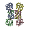
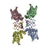
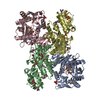
| Title | Crystal structure of SLA Reductase YihU from E. Coli in complex with NADH | ||||||
 Components Components | 3-sulfolactaldehyde reductase | ||||||
 Keywords Keywords | OXIDOREDUCTASE / apo / reductase / sulfoquinovose / dihydroxypropanesulfonate | ||||||
| Function / homology |  Function and homology information Function and homology informationsulfolactaldehyde 3-reductase / 4-hydroxybutyrate dehydrogenase / 4-hydroxybutyrate dehydrogenase activity / 3-sulfolactaldehyde reductase activity / : / 6-sulfoquinovose(1-) catabolic process / toxin catabolic process / oxidoreductase activity, acting on the CH-OH group of donors, NAD or NADP as acceptor / NAD binding / NADP binding ...sulfolactaldehyde 3-reductase / 4-hydroxybutyrate dehydrogenase / 4-hydroxybutyrate dehydrogenase activity / 3-sulfolactaldehyde reductase activity / : / 6-sulfoquinovose(1-) catabolic process / toxin catabolic process / oxidoreductase activity, acting on the CH-OH group of donors, NAD or NADP as acceptor / NAD binding / NADP binding / protein homotetramerization / protein-containing complex / identical protein binding Similarity search - Function | ||||||
| Biological species |  | ||||||
| Method |  X-RAY DIFFRACTION / SYNCHROTRON / MOLECULAR REPLACEMENT / Resolution: 1.75 Å X-RAY DIFFRACTION / SYNCHROTRON / MOLECULAR REPLACEMENT / Resolution: 1.75 Å | ||||||
 Authors Authors | Sharma, M. / Davies, G.J. | ||||||
| Funding support |  United Kingdom, 1items United Kingdom, 1items
| ||||||
 Citation Citation | Journal: Acs Catalysis / Year: 2020 Title: Dynamic Structural Changes Accompany the Production of Dihydroxypropanesulfonate by Sulfolactaldehyde Reductase Authors: Sharma, M. / Abayakoon, P. / Lingford, J.P. / Epa, R. / John, A. / Jin, Y. / Goddard-Borger, E.D. / Davies, G.J. / Williams, S.J. | ||||||
| History |
|
- Structure visualization
| Structure viewer | Molecule: MolmilJmol/JSmol |
|---|
- Downloads & links
-Download
| PDBx/mmCIF format | 6smz.cif.gz | 241.6 KB | Display | PDBx/mmCIF format |
|---|---|---|---|---|
| PDB format | pdb6smz.ent.gz | 193.5 KB | Display | PDB format |
| PDBx/mmJSON format | 6smz.json.gz | Tree view | PDBx/mmJSON format | |
| Others |  Other downloads Other downloads |
-Validation report
| Arichive directory | https://data.pdbj.org/pub/pdb/validation_reports/sm/6smzftp://data.pdbj.org/pub/pdb/validation_reports/sm/6smz | HTTPS FTP |
|---|
-Related structure data
| Related structure data |  6sm7SC  6smyC S: Starting model for refinement C: citing same article ( |
|---|---|
| Similar structure data |


-Links
 PDBj
PDBj


- Assembly
| Deposited unit | 
| ||||||||||||||||||||||||||||||||||||||||||||||||||||||||||||||||||||||||||||||||||||||||||||||||||||||||||||||||||||||||||||||||||||||||||||||||||||||
|---|---|---|---|---|---|---|---|---|---|---|---|---|---|---|---|---|---|---|---|---|---|---|---|---|---|---|---|---|---|---|---|---|---|---|---|---|---|---|---|---|---|---|---|---|---|---|---|---|---|---|---|---|---|---|---|---|---|---|---|---|---|---|---|---|---|---|---|---|---|---|---|---|---|---|---|---|---|---|---|---|---|---|---|---|---|---|---|---|---|---|---|---|---|---|---|---|---|---|---|---|---|---|---|---|---|---|---|---|---|---|---|---|---|---|---|---|---|---|---|---|---|---|---|---|---|---|---|---|---|---|---|---|---|---|---|---|---|---|---|---|---|---|---|---|---|---|---|---|---|---|---|
| 1 |
| ||||||||||||||||||||||||||||||||||||||||||||||||||||||||||||||||||||||||||||||||||||||||||||||||||||||||||||||||||||||||||||||||||||||||||||||||||||||
| Unit cell |
| ||||||||||||||||||||||||||||||||||||||||||||||||||||||||||||||||||||||||||||||||||||||||||||||||||||||||||||||||||||||||||||||||||||||||||||||||||||||
| Noncrystallographic symmetry (NCS) | NCS domain:
NCS domain segments: Component-ID: _ / Beg auth comp-ID: ALA / Beg label comp-ID: ALA / Refine code: _
NCS ensembles :
|
-Components
| #1: Protein | Mass: 32257.246 Da / Num. of mol.: 4 Source method: isolated from a genetically manipulated source Source: (gene. exp.) Strain: K12 / Gene: yihU, b3882, JW3853 / Plasmid: pET28a(+) / Production host: #2: Chemical | ChemComp-NAD /   Mass: 663.425 Da / Num. of mol.: 4 / Source method: obtained synthetically / Formula: C21H27N7O14P2 / Feature type: SUBJECT OF INVESTIGATION / Comment: NAD*YM Mass: 663.425 Da / Num. of mol.: 4 / Source method: obtained synthetically / Formula: C21H27N7O14P2 / Feature type: SUBJECT OF INVESTIGATION / Comment: NAD*YM#3: Water | ChemComp-HOH / |  Mass: 18.015 Da / Num. of mol.: 804 / Source method: isolated from a natural source / Formula: H2O Mass: 18.015 Da / Num. of mol.: 804 / Source method: isolated from a natural source / Formula: H2OHas ligand of interest | Y | |
|---|
-Experimental details
-Experiment
| Experiment | Method: X-RAY DIFFRACTION / Number of used crystals: 1 |
|---|
- Sample preparation
| Crystal | Density Matthews: 2.28 Å3/Da / Density % sol: 46.07 % |
|---|---|
| Crystal grow | Temperature: 293 K / Method: vapor diffusion, sitting drop / pH: 6.5 Details: YihU was co-crystallized with 5 mM NADH using 0.2 M Ammonium acetate, 0.1 M Bis-Tris pH 6.5, 25% w/v PEG 3,350 as precipitant. |
-Data collection
| Diffraction | Mean temperature: 100 K / Serial crystal experiment: N |
|---|---|
| Diffraction source | Source: SYNCHROTRON / Site: Diamond / Beamline: I04 / Wavelength: 0.9795 Å |
| Detector | Type: DECTRIS PILATUS 6M-F / Detector: PIXEL / Date: Jul 1, 2018 |
| Radiation | Protocol: SINGLE WAVELENGTH / Monochromatic (M) / Laue (L): M / Scattering type: x-ray |
| Radiation wavelength | Wavelength: 0.9795 Å / Relative weight: 1 |
| Reflection | Resolution: 1.75→88.95 Å / Num. obs: 119505 / % possible obs: 100 % / Redundancy: 8.2 % / CC1/2: 0.998 / Rmerge(I) obs: 0.057 / Rpim(I) all: 0.021 / Rrim(I) all: 0.061 / Net I/σ(I): 18.8 / Num. measured all: 976872 / Scaling rejects: 165 |
| Reflection shell | Resolution: 1.75→1.78 Å / Redundancy: 8.3 % / Rmerge(I) obs: 0.473 / Num. unique obs: 5864 / CC1/2: 0.954 / Rpim(I) all: 0.172 / Rrim(I) all: 0.504 / % possible all: 100 |
- Processing
| Software |
| |||||||||||||||||||||||||||||||||||||||||||||||||||||||||||||||||
|---|---|---|---|---|---|---|---|---|---|---|---|---|---|---|---|---|---|---|---|---|---|---|---|---|---|---|---|---|---|---|---|---|---|---|---|---|---|---|---|---|---|---|---|---|---|---|---|---|---|---|---|---|---|---|---|---|---|---|---|---|---|---|---|---|---|---|
| Refinement | Method to determine structure: MOLECULAR REPLACEMENT Starting model: 6SM7 Resolution: 1.75→88.95 Å / Cor.coef. Fo:Fc: 0.966 / Cor.coef. Fo:Fc free: 0.961 / SU B: 2.375 / SU ML: 0.075 / Cross valid method: THROUGHOUT / σ(F): 0 / ESU R: 0.111 / ESU R Free: 0.103 / Stereochemistry target values: MAXIMUM LIKELIHOOD Details: HYDROGENS HAVE BEEN ADDED IN THE RIDING POSITIONS U VALUES : REFINED INDIVIDUALLY
| |||||||||||||||||||||||||||||||||||||||||||||||||||||||||||||||||
| Solvent computation | Ion probe radii: 0.8 Å / Shrinkage radii: 0.8 Å / VDW probe radii: 1.2 Å / Solvent model: MASK | |||||||||||||||||||||||||||||||||||||||||||||||||||||||||||||||||
| Displacement parameters | Biso max: 82.25 Å2 / Biso mean: 27.35 Å2 / Biso min: 15.58 Å2
| |||||||||||||||||||||||||||||||||||||||||||||||||||||||||||||||||
| Refinement step | Cycle: final / Resolution: 1.75→88.95 Å
| |||||||||||||||||||||||||||||||||||||||||||||||||||||||||||||||||
| Refine LS restraints |
| |||||||||||||||||||||||||||||||||||||||||||||||||||||||||||||||||
| Refine LS restraints NCS | Refine-ID: X-RAY DIFFRACTION / Type: interatomic distance / Weight position: 0.05
| |||||||||||||||||||||||||||||||||||||||||||||||||||||||||||||||||
| LS refinement shell | Resolution: 1.75→1.795 Å / Rfactor Rfree error: 0 / Total num. of bins used: 20
|
