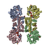
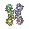
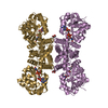
- PDB-3obb: Crystal structure of a possible 3-hydroxyisobutyrate Dehydrogenas... -
+
Open data
ID or keywords:
Loading...
-
Basic information
Entry
Database: PDB / ID: 3obb
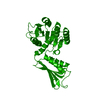
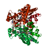
Title
Crystal structure of a possible 3-hydroxyisobutyrate Dehydrogenase from pseudomonas aeruginosa pao1
Components
Probable 3-hydroxyisobutyrate dehydrogenase
Keywords
OXIDOREDUCTASE / Structural Genomics / PSI-2 / Protein Structure Initiative / Midwest Center for Structural Genomics / MCSG / alpha-beta / Serine Dehydrogenase
Function / homology
Function and homology information
L-serine 3-dehydrogenase (NAD+) / 3-hydroxyisobutyrate dehydrogenase activity / L-serine catabolic process / branched-chain amino acid catabolic process / oxidoreductase activity, acting on the CH-OH group of donors, NAD or NADP as acceptor / NAD binding / NADP binding / protein homotetramerization Similarity search - Function
Monochromator: SI 111 CRYSTAL / Protocol: SINGLE WAVELENGTH / Monochromatic (M) / Laue (L): M / Scattering type: x-ray
Radiation wavelength
Wavelength: 0.97921 Å / Relative weight: 1
Reflection
Resolution: 2.2→30.32 Å / Num. obs: 16608 / % possible obs: 99.7 % / Observed criterion σ(I): 0 / Redundancy: 10.5 % / Rmerge(I) obs: 0.116 / Net I/σ(I): 32.6
Reflection shell
Resolution: 2.2→2.28 Å / Redundancy: 10.7 % / Rmerge(I) obs: 0.747 / Mean I/σ(I) obs: 3.56 / % possible all: 100
-
Processing
Software
Name
Version
Classification
SBC-Collect
datacollection
SHELXD
phasing
REFMAC
5.2.0019
refinement
HKL-2000
datareduction
HKL-2000
datascaling
Refinement
Method to determine structure: SAD Starting model: none Resolution: 2.2→30.32 Å / Cor.coef. Fo:Fc: 0.951 / Cor.coef. Fo:Fc free: 0.921 / SU B: 12.109 / SU ML: 0.153 / Cross valid method: THROUGHOUT / ESU R: 0.256 / ESU R Free: 0.211 / Stereochemistry target values: MAXIMUM LIKELIHOOD / Details: HYDROGENS HAVE BEEN ADDED IN THE RIDING POSITIONS
Rfactor
Num. reflection
% reflection
Selection details
Rfree
0.24774
837
5.1 %
RANDOM
Rwork
0.19653
-
-
-
obs
0.19908
15695
99.57 %
-
all
-
16532
-
-
Solvent computation
Ion probe radii: 0.8 Å / Shrinkage radii: 0.8 Å / VDW probe radii: 1.2 Å / Solvent model: MASK
Displacement parameters
Biso mean: 47.008 Å2
Baniso -1
Baniso -2
Baniso -3
1-
-0.03 Å2
-0.02 Å2
0 Å2
2-
-
-0.03 Å2
0 Å2
3-
-
-
0.05 Å2
Refinement step
Cycle: LAST / Resolution: 2.2→30.32 Å
Protein
Nucleic acid
Ligand
Solvent
Total
Num. atoms
2135
0
31
78
2244
Refine LS restraints
Refine-ID
Type
Dev ideal
Dev ideal target
Number
X-RAY DIFFRACTION
r_bond_refined_d
0.015
0.022
2229
X-RAY DIFFRACTION
r_bond_other_d
X-RAY DIFFRACTION
r_angle_refined_deg
1.649
1.986
2978
X-RAY DIFFRACTION
r_angle_other_deg
X-RAY DIFFRACTION
r_dihedral_angle_1_deg
6.974
5
295
X-RAY DIFFRACTION
r_dihedral_angle_2_deg
44.332
24.512
82
X-RAY DIFFRACTION
r_dihedral_angle_3_deg
19.724
15
350
X-RAY DIFFRACTION
r_dihedral_angle_4_deg
21.4
15
12
X-RAY DIFFRACTION
r_chiral_restr
0.115
0.2
340
X-RAY DIFFRACTION
r_gen_planes_refined
0.006
0.02
1646
X-RAY DIFFRACTION
r_gen_planes_other
X-RAY DIFFRACTION
r_nbd_refined
0.22
0.2
1086
X-RAY DIFFRACTION
r_nbd_other
X-RAY DIFFRACTION
r_nbtor_refined
0.305
0.2
1500
X-RAY DIFFRACTION
r_nbtor_other
X-RAY DIFFRACTION
r_xyhbond_nbd_refined
0.137
0.2
94
X-RAY DIFFRACTION
r_xyhbond_nbd_other
X-RAY DIFFRACTION
r_metal_ion_refined
X-RAY DIFFRACTION
r_metal_ion_other
X-RAY DIFFRACTION
r_symmetry_vdw_refined
0.212
0.2
76
X-RAY DIFFRACTION
r_symmetry_vdw_other
X-RAY DIFFRACTION
r_symmetry_hbond_refined
0.12
0.2
9
X-RAY DIFFRACTION
r_symmetry_hbond_other
X-RAY DIFFRACTION
r_symmetry_metal_ion_refined
X-RAY DIFFRACTION
r_symmetry_metal_ion_other
X-RAY DIFFRACTION
r_mcbond_it
0.805
1.5
1494
X-RAY DIFFRACTION
r_mcbond_other
X-RAY DIFFRACTION
r_mcangle_it
1.235
2
2295
X-RAY DIFFRACTION
r_scbond_it
2.382
3
782
X-RAY DIFFRACTION
r_scangle_it
3.063
4.5
682
X-RAY DIFFRACTION
r_rigid_bond_restr
X-RAY DIFFRACTION
r_sphericity_free
X-RAY DIFFRACTION
r_sphericity_bonded
LS refinement shell
Resolution: 2.203→2.26 Å / Total num. of bins used: 20
Rfactor
Num. reflection
% reflection
Rfree
0.31
57
-
Rwork
0.261
1141
-
obs
-
1142
99.34 %
Refinement TLS params.
Method: refined / Refine-ID: X-RAY DIFFRACTION
ID
L11 (°2)
L12 (°2)
L13 (°2)
L22 (°2)
L23 (°2)
L33 (°2)
S11 (Å °)
S12 (Å °)
S13 (Å °)
S21 (Å °)
S22 (Å °)
S23 (Å °)
S31 (Å °)
S32 (Å °)
S33 (Å °)
T11 (Å2)
T12 (Å2)
T13 (Å2)
T22 (Å2)
T23 (Å2)
T33 (Å2)
Origin x (Å)
Origin y (Å)
Origin z (Å)
1
2.3078
-0.127
-2.3194
0.7305
-1.3952
6.5393
-0.3273
0.4732
-0.3804
0.0018
0.1122
0.2456
1.2321
-0.6837
0.2151
0.1758
-0.2644
0.0761
-0.0345
-0.0877
-0.0068
-10.4074
25.0195
47.232
2
0.8605
0.2706
0.0183
1.241
0.0157
1.4505
-0.0413
-0.0691
0.0117
0.1511
0.111
0.0761
0.0019
-0.0219
-0.0696
-0.0616
0.0133
0.0407
-0.1088
0.0271
-0.0787
10.719
44.1632
50.3607
Refinement TLS group
ID
Refine-ID
Refine TLS-ID
Auth asym-ID
Auth seq-ID
1
X-RAY DIFFRACTION
1
A
1 - 166
2
X-RAY DIFFRACTION
2
A
167 - 296
+
About Yorodumi
-
News
-
Feb 9, 2022. New format data for meta-information of EMDB entries
New format data for meta-information of EMDB entries
Version 3 of the EMDB header file is now the official format.
The previous official version 1.9 will be removed from the archive.
In the structure databanks used in Yorodumi, some data are registered as the other names, "COVID-19 virus" and "2019-nCoV". Here are the details of the virus and the list of structure data.
Jan 31, 2019. EMDB accession codes are about to change! (news from PDBe EMDB page)
EMDB accession codes are about to change! (news from PDBe EMDB page)
The allocation of 4 digits for EMDB accession codes will soon come to an end. Whilst these codes will remain in use, new EMDB accession codes will include an additional digit and will expand incrementally as the available range of codes is exhausted. The current 4-digit format prefixed with “EMD-” (i.e. EMD-XXXX) will advance to a 5-digit format (i.e. EMD-XXXXX), and so on. It is currently estimated that the 4-digit codes will be depleted around Spring 2019, at which point the 5-digit format will come into force.
The EM Navigator/Yorodumi systems omit the EMD- prefix.
Related info.:Q: What is EMD? / ID/Accession-code notation in Yorodumi/EM Navigator
Yorodumi is a browser for structure data from EMDB, PDB, SASBDB, etc.
This page is also the successor to EM Navigator detail page, and also detail information page/front-end page for Omokage search.
The word "yorodu" (or yorozu) is an old Japanese word meaning "ten thousand". "mi" (miru) is to see.
Related info.:EMDB / PDB / SASBDB / Comparison of 3 databanks / Yorodumi Search / Aug 31, 2016. New EM Navigator & Yorodumi / Yorodumi Papers / Jmol/JSmol / Function and homology information / Changes in new EM Navigator and Yorodumi
 Movie
Movie Controller

 Yorodumi
 Open data
 Basic information
 Components
 Keywords
 Function and homology information
 Pseudomonas aeruginosa (bacteria)
 X-RAY DIFFRACTION /
  Authors
 Citation
 Structure visualization
 Downloads & links
 Other downloads
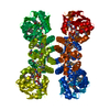


 PDBj
PDBj


 Assembly


 Mass: 59.044 Da / Num. of mol.: 3 / Source method: obtained synthetically / Formula: C2H3O2
 Mass: 62.068 Da / Num. of mol.: 1 / Source method: obtained synthetically / Formula: C2H6O2
 Mass: 238.305 Da / Num. of mol.: 1 / Source method: obtained synthetically / Formula: C8H18N2O4S / Comment: pH buffer*YM
 Mass: 18.015 Da / Num. of mol.: 78 / Source method: isolated from a natural source / Formula: H2O
 Sample preparation
 / Beamline: 19-ID / Wavelength: 0.97921 Å
 Processing