 Movie
Movie Controller


+ Open data

- Basic information
| Entry | Database: PDB / ID: 6smt | ||||||
|---|---|---|---|---|---|---|---|
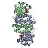
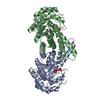
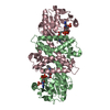
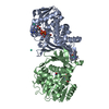
| Title | S-enantioselective imine reductase from Mycobacterium smegmatis | ||||||
 Components Components | 6-phosphogluconate dehydrogenase, NAD-binding | ||||||
 Keywords Keywords | OXIDOREDUCTASE / Imine reductase | ||||||
| Function / homology |  Function and homology information Function and homology information | ||||||
| Biological species |  Mycolicibacterium smegmatis MC2 155 (bacteria) Mycolicibacterium smegmatis MC2 155 (bacteria) | ||||||
| Method |  X-RAY DIFFRACTION / SYNCHROTRON / MOLECULAR REPLACEMENT / molecular replacement / Resolution: 1.55 Å X-RAY DIFFRACTION / SYNCHROTRON / MOLECULAR REPLACEMENT / molecular replacement / Resolution: 1.55 Å | ||||||
 Authors Authors | Meyer, T. / Zumbraegel, N. / Geerds, C. / Groeger, H. / Niemann, H.H. | ||||||
| Funding support |  Germany, 1items Germany, 1items
| ||||||
 Citation Citation | Journal: Biomolecules / Year: 2020 Title: Structural Characterization of an S -enantioselective Imine Reductase from Mycobacterium Smegmatis . Authors: Meyer, T. / Zumbragel, N. / Geerds, C. / Groger, H. / Niemann, H.H. | ||||||
| History |
|
- Structure visualization
| Structure viewer | Molecule: MolmilJmol/JSmol |
|---|
- Downloads & links
-Download
| PDBx/mmCIF format | 6smt.cif.gz | 581.5 KB | Display | PDBx/mmCIF format |
|---|---|---|---|---|
| PDB format | pdb6smt.ent.gz | 479.6 KB | Display | PDB format |
| PDBx/mmJSON format | 6smt.json.gz | Tree view | PDBx/mmJSON format | |
| Others |  Other downloads Other downloads |
-Validation report
| Arichive directory | https://data.pdbj.org/pub/pdb/validation_reports/sm/6smtftp://data.pdbj.org/pub/pdb/validation_reports/sm/6smt | HTTPS FTP |
|---|
-Related structure data


-Links
 PDBj
PDBj


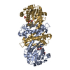
- Assembly
| Deposited unit | 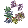
| ||||||||
|---|---|---|---|---|---|---|---|---|---|
| 1 | 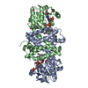
| ||||||||
| 2 | 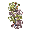
| ||||||||
| 3 | 
| ||||||||
| Unit cell |
|
-Components
-Protein , 1 types, 5 molecules ABCDE
| #1: Protein | Mass: 31034.783 Da / Num. of mol.: 5 Source method: isolated from a genetically manipulated source Source: (gene. exp.) Mycolicibacterium smegmatis MC2 155 (bacteria)Gene: MSMEG_6341 / Production host: |
|---|
-Non-polymers , 7 types, 1999 molecules 


| #2: Chemical | ChemComp-NDP /  Mass: 745.421 Da / Num. of mol.: 5 / Source method: obtained synthetically / Formula: C21H30N7O17P3 Mass: 745.421 Da / Num. of mol.: 5 / Source method: obtained synthetically / Formula: C21H30N7O17P3#3: Chemical | ChemComp-FMT /  Mass: 46.025 Da / Num. of mol.: 12 / Source method: obtained synthetically / Formula: CH2O2 Mass: 46.025 Da / Num. of mol.: 12 / Source method: obtained synthetically / Formula: CH2O2#4: Chemical |  Mass: 62.068 Da / Num. of mol.: 3 / Source method: obtained synthetically / Formula: C2H6O2 Mass: 62.068 Da / Num. of mol.: 3 / Source method: obtained synthetically / Formula: C2H6O2#5: Chemical | ChemComp-2EH / (  Mass: 130.228 Da / Num. of mol.: 5 / Source method: obtained synthetically / Formula: C8H18O Mass: 130.228 Da / Num. of mol.: 5 / Source method: obtained synthetically / Formula: C8H18O#6: Chemical |  Mass: 92.094 Da / Num. of mol.: 3 / Source method: obtained synthetically / Formula: C3H8O3 Mass: 92.094 Da / Num. of mol.: 3 / Source method: obtained synthetically / Formula: C3H8O3#7: Chemical | ChemComp-PEG / |  Mass: 106.120 Da / Num. of mol.: 1 / Source method: obtained synthetically / Formula: C4H10O3 Mass: 106.120 Da / Num. of mol.: 1 / Source method: obtained synthetically / Formula: C4H10O3#8: Water | ChemComp-HOH / | Mass: 18.015 Da / Num. of mol.: 1970 / Source method: isolated from a natural source / Formula: H2O |
|---|
-Details
| Has ligand of interest | N |
|---|
-Experimental details
-Experiment
| Experiment | Method: X-RAY DIFFRACTION / Number of used crystals: 1 |
|---|
- Sample preparation
| Crystal | Density Matthews: 2.96 Å3/Da / Density % sol: 58.49 % |
|---|---|
| Crystal grow | Temperature: 277.15 K / Method: vapor diffusion, sitting drop / Details: 20 % (w/v) PEG 3350, 0.2 M sodium formate |
-Data collection
| Diffraction | Mean temperature: 100 K / Serial crystal experiment: N | ||||||||||||||||||||||||||||||
|---|---|---|---|---|---|---|---|---|---|---|---|---|---|---|---|---|---|---|---|---|---|---|---|---|---|---|---|---|---|---|---|
| Diffraction source | Source: SYNCHROTRON / Site: PETRA III, EMBL c/o DESY / Beamline: P13 (MX1) / Wavelength: 0.9762 Å | ||||||||||||||||||||||||||||||
| Detector | Type: DECTRIS PILATUS 6M-F / Detector: PIXEL / Date: Sep 18, 2018 | ||||||||||||||||||||||||||||||
| Radiation | Protocol: SINGLE WAVELENGTH / Monochromatic (M) / Laue (L): M / Scattering type: x-ray | ||||||||||||||||||||||||||||||
| Radiation wavelength | Wavelength: 0.9762 Å / Relative weight: 1 | ||||||||||||||||||||||||||||||
| Reflection | Resolution: 1.55→100.42 Å / Num. obs: 248023 / % possible obs: 99.1 % / Redundancy: 6.9 % / CC1/2: 0.997 / Rmerge(I) obs: 0.073 / Rpim(I) all: 0.03 / Rrim(I) all: 0.08 / Net I/σ(I): 13.2 / Num. measured all: 1702995 / Scaling rejects: 6558 | ||||||||||||||||||||||||||||||
| Reflection shell | Diffraction-ID: 1
|
-Phasing
| Phasing | Method: molecular replacement |
|---|
- Processing
| Software |
| ||||||||||||||||||||||||||||||||||||||||||||||||||||||||||||||||||||||||||||||||||||||||||||||||||||||||||||||||||||||||||||||||||||||||||||||||||||||
|---|---|---|---|---|---|---|---|---|---|---|---|---|---|---|---|---|---|---|---|---|---|---|---|---|---|---|---|---|---|---|---|---|---|---|---|---|---|---|---|---|---|---|---|---|---|---|---|---|---|---|---|---|---|---|---|---|---|---|---|---|---|---|---|---|---|---|---|---|---|---|---|---|---|---|---|---|---|---|---|---|---|---|---|---|---|---|---|---|---|---|---|---|---|---|---|---|---|---|---|---|---|---|---|---|---|---|---|---|---|---|---|---|---|---|---|---|---|---|---|---|---|---|---|---|---|---|---|---|---|---|---|---|---|---|---|---|---|---|---|---|---|---|---|---|---|---|---|---|---|---|---|
| Refinement | Method to determine structure: MOLECULAR REPLACEMENT Starting model: 5G6R, 3ZGY, 4OQY, 4D3S Resolution: 1.55→100.42 Å / Cor.coef. Fo:Fc: 0.979 / Cor.coef. Fo:Fc free: 0.97 / SU B: 1.811 / SU ML: 0.036 / Cross valid method: THROUGHOUT / σ(F): 0 / ESU R: 0.056 / ESU R Free: 0.058 / Stereochemistry target values: MAXIMUM LIKELIHOOD Details: HYDROGENS HAVE BEEN ADDED IN THE RIDING POSITIONS U VALUES : WITH TLS ADDED
| ||||||||||||||||||||||||||||||||||||||||||||||||||||||||||||||||||||||||||||||||||||||||||||||||||||||||||||||||||||||||||||||||||||||||||||||||||||||
| Solvent computation | Ion probe radii: 0.8 Å / Shrinkage radii: 0.8 Å / VDW probe radii: 1.2 Å / Solvent model: MASK | ||||||||||||||||||||||||||||||||||||||||||||||||||||||||||||||||||||||||||||||||||||||||||||||||||||||||||||||||||||||||||||||||||||||||||||||||||||||
| Displacement parameters | Biso max: 69.91 Å2 / Biso mean: 18.425 Å2 / Biso min: 9.01 Å2
| ||||||||||||||||||||||||||||||||||||||||||||||||||||||||||||||||||||||||||||||||||||||||||||||||||||||||||||||||||||||||||||||||||||||||||||||||||||||
| Refinement step | Cycle: final / Resolution: 1.55→100.42 Å
| ||||||||||||||||||||||||||||||||||||||||||||||||||||||||||||||||||||||||||||||||||||||||||||||||||||||||||||||||||||||||||||||||||||||||||||||||||||||
| Refine LS restraints |
| ||||||||||||||||||||||||||||||||||||||||||||||||||||||||||||||||||||||||||||||||||||||||||||||||||||||||||||||||||||||||||||||||||||||||||||||||||||||
| LS refinement shell | Resolution: 1.55→1.59 Å / Rfactor Rfree error: 0 / Total num. of bins used: 20
| ||||||||||||||||||||||||||||||||||||||||||||||||||||||||||||||||||||||||||||||||||||||||||||||||||||||||||||||||||||||||||||||||||||||||||||||||||||||
| Refinement TLS params. | Method: refined / Refine-ID: X-RAY DIFFRACTION
| ||||||||||||||||||||||||||||||||||||||||||||||||||||||||||||||||||||||||||||||||||||||||||||||||||||||||||||||||||||||||||||||||||||||||||||||||||||||
| Refinement TLS group |
|
