 Movie
Movie Controller

[English] 日本語
 Yorodumi
- PDB-6sk8: DeltaC3 C-terminal truncation of HsNMT1 in complex with MyrCoA an... -
+ Open data

- Basic information
| Entry | Database: PDB / ID: 6sk8 | |||||||||
|---|---|---|---|---|---|---|---|---|---|---|
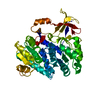
| Title | DeltaC3 C-terminal truncation of HsNMT1 in complex with MyrCoA and GDCFSKPR substrates | |||||||||
 Components Components |
| |||||||||
 Keywords Keywords | TRANSFERASE / NMT / MYRISTOYLTRANSFERASE TYPE1 / ACYLTRANSFERASE / GNAT / GCN5-RELATED N-ACETYLTRANSFERASES | |||||||||
| Function / homology |  Function and homology information Function and homology informationmyristoyltransferase activity / N-terminal peptidyl-glycine N-myristoylation / peptidyl-lysine N6-myristoyltransferase activity / Late Phase of HIV Life Cycle / ketone metabolic process / regulation of opsin-mediated signaling pathway / Activation, myristolyation of BID and translocation to mitochondria / positive regulation of protein localization to mitochondrion / glycylpeptide N-tetradecanoyltransferase / glycylpeptide N-tetradecanoyltransferase activity ...myristoyltransferase activity / N-terminal peptidyl-glycine N-myristoylation / peptidyl-lysine N6-myristoyltransferase activity / Late Phase of HIV Life Cycle / ketone metabolic process / regulation of opsin-mediated signaling pathway / Activation, myristolyation of BID and translocation to mitochondria / positive regulation of protein localization to mitochondrion / glycylpeptide N-tetradecanoyltransferase / glycylpeptide N-tetradecanoyltransferase activity / Oxidoreductases / protein localization to membrane / execution phase of apoptosis / oxidoreductase activity, acting on NAD(P)H / eNOS activation / Transferases; Acyltransferases; Transferring groups other than aminoacyl groups / 2 iron, 2 sulfur cluster binding / Inactivation, recovery and regulation of the phototransduction cascade / in utero embryonic development / mitochondrial inner membrane / endoplasmic reticulum / mitochondrion / metal ion binding / plasma membrane / cytoplasm / cytosol Similarity search - Function | |||||||||
| Biological species |  Homo sapiens (human) Homo sapiens (human) | |||||||||
| Method |  X-RAY DIFFRACTION / SYNCHROTRON / MOLECULAR REPLACEMENT / molecular replacement / Resolution: 1.87 Å X-RAY DIFFRACTION / SYNCHROTRON / MOLECULAR REPLACEMENT / molecular replacement / Resolution: 1.87 Å | |||||||||
 Authors Authors | Dian, C. / Riviere, F.B. / Asensio, T. / Giglione, C. / Meinnel, T. | |||||||||
| Funding support |  France, 2items France, 2items
| |||||||||
 Citation Citation | Journal: Nat Commun / Year: 2020 Title: High-resolution snapshots of human N-myristoyltransferase in action illuminate a mechanism promoting N-terminal Lys and Gly myristoylation. Authors: Dian, C. / Perez-Dorado, I. / Riviere, F. / Asensio, T. / Legrand, P. / Ritzefeld, M. / Shen, M. / Cota, E. / Meinnel, T. / Tate, E.W. / Giglione, C. | |||||||||
| History |
|
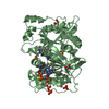
- Structure visualization
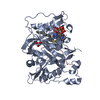
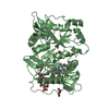
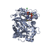
| Structure viewer | Molecule: MolmilJmol/JSmol |
|---|
- Downloads & links
-Download
| PDBx/mmCIF format | 6sk8.cif.gz | 201.8 KB | Display | PDBx/mmCIF format |
|---|---|---|---|---|
| PDB format | pdb6sk8.ent.gz | 156.7 KB | Display | PDB format |
| PDBx/mmJSON format | 6sk8.json.gz | Tree view | PDBx/mmJSON format | |
| Others |  Other downloads Other downloads |
-Validation report
| Arichive directory | https://data.pdbj.org/pub/pdb/validation_reports/sk/6sk8ftp://data.pdbj.org/pub/pdb/validation_reports/sk/6sk8 | HTTPS FTP |
|---|
-Related structure data
| Related structure data |  6ehjC  6qrmC 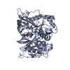 6sjzC 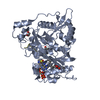 6sk2C 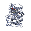 6sk3C  6skjC 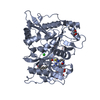 5o9vS S: Starting model for refinement C: citing same article ( |
|---|---|
| Similar structure data |


-Links
 PDBj
PDBj


- Assembly
| Deposited unit | 
| ||||||||
|---|---|---|---|---|---|---|---|---|---|
| 1 | 
| ||||||||
| 2 | 
| ||||||||
| Unit cell |
|
-Components
-Protein / Protein/peptide , 2 types, 4 molecules ABCD
| #1: Protein | Mass: 46124.996 Da / Num. of mol.: 2 / Mutation: V494stop Source method: isolated from a genetically manipulated source Source: (gene. exp.) Homo sapiens (human) / Gene: NMT1, NMT / Plasmid: pET28 derivative / Production host:  References: UniProt: P30419, glycylpeptide N-tetradecanoyltransferase #2: Protein/peptide | Mass: 911.038 Da / Num. of mol.: 2 / Mutation: G3D, P8R / Source method: obtained synthetically / Source: (synth.) Homo sapiens (human) / References: UniProt: Q96NN9*PLUS |
|---|
-Non-polymers , 4 types, 852 molecules 


| #3: Chemical |  Mass: 977.890 Da / Num. of mol.: 2 / Source method: obtained synthetically / Formula: C35H62N7O17P3S / Feature type: SUBJECT OF INVESTIGATION Mass: 977.890 Da / Num. of mol.: 2 / Source method: obtained synthetically / Formula: C35H62N7O17P3S / Feature type: SUBJECT OF INVESTIGATION#4: Chemical | ChemComp-GOL /  Mass: 92.094 Da / Num. of mol.: 6 / Source method: obtained synthetically / Formula: C3H8O3 Mass: 92.094 Da / Num. of mol.: 6 / Source method: obtained synthetically / Formula: C3H8O3#5: Chemical | ChemComp-CL / |  Mass: 35.453 Da / Num. of mol.: 1 / Source method: obtained synthetically / Formula: Cl Mass: 35.453 Da / Num. of mol.: 1 / Source method: obtained synthetically / Formula: Cl#6: Water | ChemComp-HOH / | Mass: 18.015 Da / Num. of mol.: 843 / Source method: isolated from a natural source / Formula: H2O |
|---|
-Details
| Has ligand of interest | Y |
|---|
-Experimental details
-Experiment
| Experiment | Method: X-RAY DIFFRACTION / Number of used crystals: 1 |
|---|
- Sample preparation
| Crystal | Density Matthews: 2.22 Å3/Da / Density % sol: 44.57 % |
|---|---|
| Crystal grow | Temperature: 293 K / Method: vapor diffusion, hanging drop / pH: 5.6 Details: 19% PEG8K, 100mM Sodium Citrate pH 5.6, 100mM MgCl2, 100mM NaCl |
-Data collection
| Diffraction | Mean temperature: 100 K / Ambient temp details: 100 K cryostream / Serial crystal experiment: N | ||||||||||||||||||||||||||||||
|---|---|---|---|---|---|---|---|---|---|---|---|---|---|---|---|---|---|---|---|---|---|---|---|---|---|---|---|---|---|---|---|
| Diffraction source | Source: SYNCHROTRON / Site: SOLEIL / Beamline: PROXIMA 1 / Wavelength: 0.978565 Å | ||||||||||||||||||||||||||||||
| Detector | Type: DECTRIS EIGER X 16M / Detector: PIXEL / Date: Jul 6, 2019 | ||||||||||||||||||||||||||||||
| Radiation | Monochromator: Si (111) crystal / Protocol: SINGLE WAVELENGTH / Monochromatic (M) / Laue (L): M / Scattering type: x-ray | ||||||||||||||||||||||||||||||
| Radiation wavelength | Wavelength: 0.978565 Å / Relative weight: 1 | ||||||||||||||||||||||||||||||
| Reflection | Resolution: 1.87→49.31 Å / Num. obs: 68387 / % possible obs: 99.9 % / Redundancy: 7.4 % / Biso Wilson estimate: 25.54 Å2 / CC1/2: 0.994 / Rmerge(I) obs: 0.159 / Rpim(I) all: 0.063 / Rrim(I) all: 0.171 / Net I/σ(I): 7.3 / Num. measured all: 507596 | ||||||||||||||||||||||||||||||
| Reflection shell | Diffraction-ID: 1
|
-Phasing
| Phasing | Method: molecular replacement | |||||||||
|---|---|---|---|---|---|---|---|---|---|---|
| Phasing MR |
|
- Processing
| Software |
| |||||||||||||||||||||||||||||||||||||||||||||||||||||||||||||||||||||||||||||||||||||||||||||||||||||||||||||||||||||||||||||
|---|---|---|---|---|---|---|---|---|---|---|---|---|---|---|---|---|---|---|---|---|---|---|---|---|---|---|---|---|---|---|---|---|---|---|---|---|---|---|---|---|---|---|---|---|---|---|---|---|---|---|---|---|---|---|---|---|---|---|---|---|---|---|---|---|---|---|---|---|---|---|---|---|---|---|---|---|---|---|---|---|---|---|---|---|---|---|---|---|---|---|---|---|---|---|---|---|---|---|---|---|---|---|---|---|---|---|---|---|---|---|---|---|---|---|---|---|---|---|---|---|---|---|---|---|---|---|
| Refinement | Method to determine structure: MOLECULAR REPLACEMENT Starting model: 5O9V Resolution: 1.87→49.31 Å / SU ML: 0.27 / Cross valid method: THROUGHOUT / σ(F): 1.34 / Phase error: 28.52
| |||||||||||||||||||||||||||||||||||||||||||||||||||||||||||||||||||||||||||||||||||||||||||||||||||||||||||||||||||||||||||||
| Solvent computation | Shrinkage radii: 0.9 Å / VDW probe radii: 1.11 Å | |||||||||||||||||||||||||||||||||||||||||||||||||||||||||||||||||||||||||||||||||||||||||||||||||||||||||||||||||||||||||||||
| Displacement parameters | Biso max: 85.35 Å2 / Biso mean: 30.2305 Å2 / Biso min: 14.35 Å2 | |||||||||||||||||||||||||||||||||||||||||||||||||||||||||||||||||||||||||||||||||||||||||||||||||||||||||||||||||||||||||||||
| Refinement step | Cycle: final / Resolution: 1.87→49.31 Å
| |||||||||||||||||||||||||||||||||||||||||||||||||||||||||||||||||||||||||||||||||||||||||||||||||||||||||||||||||||||||||||||
| Refine LS restraints |
| |||||||||||||||||||||||||||||||||||||||||||||||||||||||||||||||||||||||||||||||||||||||||||||||||||||||||||||||||||||||||||||
| LS refinement shell | Refine-ID: X-RAY DIFFRACTION / Rfactor Rfree error: 0 / % reflection obs: 100 %
|
