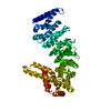
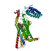
 Movie
Movie Controller


+ Open data

- Basic information
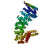
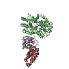
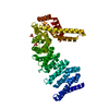
| Entry | Database: PDB / ID: 6shc | |||||||||
|---|---|---|---|---|---|---|---|---|---|---|
| Title | Crystal structure of human IRE1 luminal domain Q105C | |||||||||
 Components Components | Serine/threonine-protein kinase/endoribonuclease IRE1 | |||||||||
 Keywords Keywords | SIGNALING PROTEIN / IRE1 / unfolded protein response / ER stress / protein quality control | |||||||||
| Function / homology |  Function and homology information Function and homology informationAIP1-IRE1 complex / Ire1 complex / mRNA splicing, via endonucleolytic cleavage and ligation / IRE1alpha activates chaperones / IRE1-TRAF2-ASK1 complex / insulin metabolic process / positive regulation of endoplasmic reticulum unfolded protein response / Hydrolases; Acting on ester bonds; Endoribonucleases producing 5'-phosphomonoesters / platelet-derived growth factor receptor binding / endothelial cell proliferation ...AIP1-IRE1 complex / Ire1 complex / mRNA splicing, via endonucleolytic cleavage and ligation / IRE1alpha activates chaperones / IRE1-TRAF2-ASK1 complex / insulin metabolic process / positive regulation of endoplasmic reticulum unfolded protein response / Hydrolases; Acting on ester bonds; Endoribonucleases producing 5'-phosphomonoesters / platelet-derived growth factor receptor binding / endothelial cell proliferation / nuclear inner membrane / IRE1-RACK1-PP2A complex / IRE1-mediated unfolded protein response / mRNA catabolic process / negative regulation of intrinsic apoptotic signaling pathway / cellular response to vascular endothelial growth factor stimulus / intrinsic apoptotic signaling pathway in response to endoplasmic reticulum stress / cellular response to unfolded protein / regulation of macroautophagy / positive regulation of vascular associated smooth muscle cell proliferation / endoplasmic reticulum unfolded protein response / RNA endonuclease activity / Hsp70 protein binding / response to endoplasmic reticulum stress / positive regulation of RNA splicing / cellular response to glucose stimulus / Hsp90 protein binding / ADP binding / positive regulation of JNK cascade / cellular response to hydrogen peroxide / : / protein phosphorylation / non-specific serine/threonine protein kinase / protein serine kinase activity / hydrolase activity / protein serine/threonine kinase activity / endoplasmic reticulum membrane / magnesium ion binding / enzyme binding / endoplasmic reticulum / protein homodimerization activity / mitochondrion / ATP binding / identical protein binding / cytoplasm Similarity search - Function | |||||||||
| Biological species |  Homo sapiens (human) Homo sapiens (human) | |||||||||
| Method |  X-RAY DIFFRACTION / SYNCHROTRON / MOLECULAR REPLACEMENT / Resolution: 3.55 Å X-RAY DIFFRACTION / SYNCHROTRON / MOLECULAR REPLACEMENT / Resolution: 3.55 Å | |||||||||
 Authors Authors | Yan, Y. / Ron, D. | |||||||||
| Funding support |  United Kingdom, 2items United Kingdom, 2items
| |||||||||
 Citation Citation | Journal: Elife / Year: 2019 Title: Unstructured regions in IRE1 alpha specify BiP-mediated destabilisation of the luminal domain dimer and repression of the UPR. Authors: Amin-Wetzel, N. / Neidhardt, L. / Yan, Y. / Mayer, M.P. / Ron, D. | |||||||||
| History |
|
- Structure visualization
| Structure viewer | Molecule: MolmilJmol/JSmol |
|---|
- Downloads & links
-Download
| PDBx/mmCIF format | 6shc.cif.gz | 72.4 KB | Display | PDBx/mmCIF format |
|---|---|---|---|---|
| PDB format | pdb6shc.ent.gz | 42.7 KB | Display | PDB format |
| PDBx/mmJSON format | 6shc.json.gz | Tree view | PDBx/mmJSON format | |
| Others |  Other downloads Other downloads |
-Validation report
| Arichive directory | https://data.pdbj.org/pub/pdb/validation_reports/sh/6shcftp://data.pdbj.org/pub/pdb/validation_reports/sh/6shc | HTTPS FTP |
|---|
-Related structure data
| Related structure data | 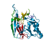 2hz6S S: Starting model for refinement |
|---|---|
| Similar structure data |


-Links
 PDBj
PDBj- Assembly
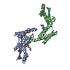
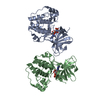
| Deposited unit | 
| ||||||||||||
|---|---|---|---|---|---|---|---|---|---|---|---|---|---|
| 1 | 
| ||||||||||||
| Unit cell |
|
-Components
| #1: Protein | Mass: 41006.367 Da / Num. of mol.: 1 / Mutation: Q105C, C109S, C148S, C332S Source method: isolated from a genetically manipulated source Details: Four mutations has been introduced into the construct: Q105C, C109S, C148S, C332S. Source: (gene. exp.) Homo sapiens (human) / Gene: ERN1, IRE1 / Production host:  References: UniProt: O75460, non-specific serine/threonine protein kinase, Hydrolases; Acting on ester bonds; Endoribonucleases producing 5'-phosphomonoesters |
|---|---|
| Has protein modification | Y |
-Experimental details
-Experiment
| Experiment | Method: X-RAY DIFFRACTION / Number of used crystals: 1 |
|---|
- Sample preparation
| Crystal | Density Matthews: 5.34 Å3/Da / Density % sol: 76.97 % |
|---|---|
| Crystal grow | Temperature: 293 K / Method: vapor diffusion / pH: 7.5 / Details: 9% MPD, 0.1M Hepes pH7.5 |
-Data collection
| Diffraction | Mean temperature: 100 K / Serial crystal experiment: N |
|---|---|
| Diffraction source | Source: SYNCHROTRON / Site: Diamond / Beamline: I04-1 / Wavelength: 0.916 Å |
| Detector | Type: DECTRIS PILATUS 6M-F / Detector: PIXEL / Date: Mar 4, 2018 |
| Radiation | Protocol: SINGLE WAVELENGTH / Monochromatic (M) / Laue (L): M / Scattering type: x-ray |
| Radiation wavelength | Wavelength: 0.916 Å / Relative weight: 1 |
| Reflection | Resolution: 3.55→91.39 Å / Num. obs: 8590 / % possible obs: 100 % / Redundancy: 19.3 % / Biso Wilson estimate: 143.03 Å2 / CC1/2: 1 / Rmerge(I) obs: 0.18 / Rpim(I) all: 0.058 / Rrim(I) all: 0.189 / Net I/σ(I): 11.7 |
| Reflection shell | Resolution: 3.55→3.89 Å / Rmerge(I) obs: 2.242 / Mean I/σ(I) obs: 1.5 / Num. unique obs: 1996 / CC1/2: 0.797 / Rpim(I) all: 0.719 / Rrim(I) all: 2.355 |
- Processing
| Software |
| ||||||||||||||||||||||||||||
|---|---|---|---|---|---|---|---|---|---|---|---|---|---|---|---|---|---|---|---|---|---|---|---|---|---|---|---|---|---|
| Refinement | Method to determine structure: MOLECULAR REPLACEMENT Starting model: 2hz6 Resolution: 3.55→54.79 Å / SU ML: 0.6921 / Cross valid method: FREE R-VALUE / σ(F): 1.34 / Phase error: 42.6377 Stereochemistry target values: GeoStd + Monomer Library + CDL v1.2
| ||||||||||||||||||||||||||||
| Solvent computation | Shrinkage radii: 0.9 Å / VDW probe radii: 1.11 Å / Solvent model: FLAT BULK SOLVENT MODEL | ||||||||||||||||||||||||||||
| Displacement parameters | Biso mean: 126.62 Å2 | ||||||||||||||||||||||||||||
| Refinement step | Cycle: LAST / Resolution: 3.55→54.79 Å
| ||||||||||||||||||||||||||||
| Refine LS restraints |
| ||||||||||||||||||||||||||||
| LS refinement shell |
|
