[English] 日本語
Yorodumi
- PDB-6sa9: Endogenous Retrovirus HML2 Capsid NTD -

+
Open data


ID or keywords:

Loading...

-
Basic information

Entry
Database: PDB / ID: 6sa9
TitleEndogenous Retrovirus HML2 Capsid NTD
ComponentsEndogenous retrovirus group K member 9 Pol protein
KeywordsVIRUS LIKE PARTICLE / HML2 HERV-K Endogenous Retrovirus Capsid CA
Function / homology
Function and homology information


human endogenous retrovirus K endopeptidase / ribonuclease H / viral process / RNA-directed DNA polymerase / RNA-directed DNA polymerase activity / RNA-DNA hybrid ribonuclease activity / DNA-directed DNA polymerase / aspartic-type endopeptidase activity / DNA-directed DNA polymerase activity / structural molecule activity ...human endogenous retrovirus K endopeptidase / ribonuclease H / viral process / RNA-directed DNA polymerase / RNA-directed DNA polymerase activity / RNA-DNA hybrid ribonuclease activity / DNA-directed DNA polymerase / aspartic-type endopeptidase activity / DNA-directed DNA polymerase activity / structural molecule activity / RNA binding / zinc ion binding / plasma membrane
Similarity search - Function
Beta-retroviral matrix protein / Beta-retroviral matrix superfamily / Retroviral GAG p10 protein / Retroviral nucleocapsid Gag protein p24, N-terminal / G-patch domain / G-patch domain profile. / G-patch domain / glycine rich nucleic binding domain / dUTPase-like / dUTPase ...Beta-retroviral matrix protein / Beta-retroviral matrix superfamily / Retroviral GAG p10 protein / Retroviral nucleocapsid Gag protein p24, N-terminal / G-patch domain / G-patch domain profile. / G-patch domain / glycine rich nucleic binding domain / dUTPase-like / dUTPase / dUTPase, trimeric / dUTPase-like superfamily / gag protein p24 N-terminal domain / Retropepsin-like catalytic domain / Reverse transcriptase (RNA-dependent DNA polymerase) / Reverse transcriptase domain / Reverse transcriptase (RT) catalytic domain profile. / Retropepsins / Retroviral aspartyl protease / Aspartyl protease, retroviral-type family profile. / Peptidase A2A, retrovirus, catalytic / Retrovirus capsid, C-terminal / Retroviral matrix protein / Retrovirus capsid, N-terminal / zinc finger / Zinc knuckle / Zinc finger, CCHC-type superfamily / Zinc finger, CCHC-type / Zinc finger CCHC-type profile. / Aspartic peptidase, active site / Eukaryotic and viral aspartyl proteases active site. / Aspartic peptidase domain superfamily / Reverse transcriptase/Diguanylate cyclase domain / DNA/RNA polymerase superfamily
Similarity search - Domain/homology
Endogenous retrovirus group K member 9 Pol protein
Similarity search - Component
Biological speciesHomo sapiens (human)
MethodX-RAY DIFFRACTION / MOLECULAR REPLACEMENT / molecular replacement / Resolution: 1.8 Å
AuthorsGoldstone, D.C. / Ball, N.J. / Taylor, I.A.
Funding support United Kingdom, 1items
OrganizationGrant numberCountry
The Francis Crick Institute United Kingdom
Citation
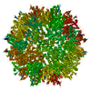
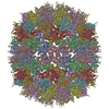
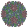
Journal: Nat Commun / Year: 2019
Title: Structural basis for Fullerene geometry in a human endogenous retrovirus capsid.
Authors: Oliver Acton / Tim Grant / Giuseppe Nicastro / Neil J Ball / David C Goldstone / Laura E Robertson / Kasim Sader / Andrea Nans / Andres Ramos / Jonathan P Stoye / Ian A Taylor / Peter B Rosenthal /
Abstract: The HML2 (HERV-K) group constitutes the most recently acquired family of human endogenous retroviruses, with many proviruses less than one million years old. Many maintain intact open reading frames ...The HML2 (HERV-K) group constitutes the most recently acquired family of human endogenous retroviruses, with many proviruses less than one million years old. Many maintain intact open reading frames and provirus expression together with HML2 particle formation are observed in early stage human embryo development and are associated with pluripotency as well as inflammatory disease, cancers and HIV-1 infection. Here, we reconstruct the core structural protein (CA) of an HML2 retrovirus, assemble particles in vitro and employ single particle cryogenic electron microscopy (cryo-EM) to determine structures of four classes of CA Fullerene shell assemblies. These icosahedral and capsular assemblies reveal at high-resolution the molecular interactions that allow CA to form both pentamers and hexamers and show how invariant pentamers and structurally plastic hexamers associate to form the unique polyhedral structures found in retroviral cores.
History
DepositionJul 16, 2019Deposition site: PDBE / Processing site: PDBE
Revision 1.0Jan 1, 2020Provider: repository / Type: Initial release

-
Structure visualization

Structure viewerMolecule:
MolmilJmol/JSmol

Downloads & links

-
Assembly

Deposited unit
A: Endogenous retrovirus group K member 9 Pol protein
B: Endogenous retrovirus group K member 9 Pol protein
hetero molecules


Theoretical massNumber of molelcules
Total (without water)37,4133
Polymers37,3202
Non-polymers921
Water5,585310
1


  • Idetical with deposited unit
  • defined by author&software
  • Evidence: equilibrium centrifugation
TypeNameSymmetry operationNumber
identity operation1_555x,y,z1
Buried area1760 Å2
ΔGint-9 kcal/mol
Surface area13900 Å2
MethodPISA
Unit cell
Length a, b, c (Å)42.387, 98.920, 76.508
Angle α, β, γ (deg.)90.000, 94.370, 90.000
Int Tables number5
Space group name H-MC121
Components on special symmetry positions
IDModelComponents
11A-402-

HOH

21B-257-

HOH

-
Components

#1: Protein Endogenous retrovirus group K member 9 Pol protein / HERV-K(C6) Gag-Pol protein / HERV-K109 Gag-Pol protein / HERV-K_6q14.1 provirus ancestral Gag-Pol polyprotein


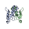
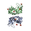
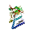
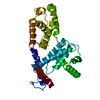
Mass: 18660.223 Da / Num. of mol.: 2
Source method: isolated from a genetically manipulated source
Source: (gene. exp.) Homo sapiens (human) / Gene: ERVK-9 / Production host: Escherichia coli (E. coli)
References: UniProt: P63128, human endogenous retrovirus K endopeptidase, RNA-directed DNA polymerase, DNA-directed DNA polymerase, ribonuclease H
#2: Chemical ChemComp-GOL / GLYCEROL / GLYCERIN / PROPANE-1,2,3-TRIOL / Glycerol


Mass: 92.094 Da / Num. of mol.: 1 / Source method: obtained synthetically / Formula: C3H8O3
#3: Water ChemComp-HOH / water / Water


Mass: 18.015 Da / Num. of mol.: 310 / Source method: isolated from a natural source / Formula: H2O
Has ligand of interestN

-
Experimental details

-
Experiment

ExperimentMethod: X-RAY DIFFRACTION / Number of used crystals: 1

-
Sample preparation

CrystalDensity Matthews: 2.54 Å3/Da / Density % sol: 51.65 %
Crystal growTemperature: 291 K / Method: vapor diffusion
Details: 0.2 M Sodium Acetate Trihydrate, 0.1 M Tris Hydrochloride and 15% (w/v) PEG 4000

-
Data collection

DiffractionMean temperature: 100 K / Serial crystal experiment: N
Diffraction sourceSource: ROTATING ANODE / Type: RIGAKU R-AXIS IV / Wavelength: 1.5418 Å
DetectorType: RIGAKU RAXIS IV / Detector: IMAGE PLATE / Date: Mar 6, 2009
RadiationProtocol: SINGLE WAVELENGTH / Monochromatic (M) / Laue (L): M / Scattering type: x-ray
Radiation wavelengthWavelength: 1.5418 Å / Relative weight: 1
ReflectionResolution: 1.8→35 Å / Num. obs: 26836 / % possible obs: 92.2 % / Redundancy: 7.1 % / Biso Wilson estimate: 21.38 Å2 / Rmerge(I) obs: 0.05 / Χ2: 1.154 / Net I/σ(I): 27.1 / Num. measured all: 191173
Reflection shell
Resolution (Å)Redundancy (%)Rmerge(I) obsNum. unique obsΧ2Diffraction-ID% possible all
1.8-1.866.50.24520661.187171.4
1.86-1.9470.19526551.192191.1
1.94-2.037.20.13826431.185191.9
2.03-2.137.20.10527041.176192.7
2.13-2.277.20.08626901.171193.2
2.27-2.447.20.0727771.118194.4
2.44-2.697.20.05827571.094195.1
2.69-3.087.20.0527951.26196.1
3.08-3.887.20.04128451.16197.3
3.88-357.10.03429041.015198.2

-
Phasing

PhasingMethod: molecular replacement
Phasing MRRfactor: 47.31 / Model details: Phaser MODE: MR_AUTO
Highest resolutionLowest resolution
Rotation2.5 Å24.93 Å
Translation2.5 Å24.93 Å

-
Processing

Software
NameVersionClassificationNB
DENZOdata reduction
SCALEPACKdata scaling
PHASER2.1.4phasing
PHENIX(phenix.refine)refinement
PDB_EXTRACT3.25data extraction
RefinementMethod to determine structure: MOLECULAR REPLACEMENT / Resolution: 1.8→24.73 Å / Cross valid method: THROUGHOUT / σ(F): 1.36 / Phase error: 18.26
RfactorNum. reflection% reflection
Rfree0.1951 1367 5.1 %
Rwork0.157 --
obs0.1589 26828 92.16 %
Solvent computationShrinkage radii: 0.9 Å / VDW probe radii: 1.11 Å / Bsol: 58.685 Å2 / ksol: 0.358 e/Å3
Displacement parametersBiso max: 95.65 Å2 / Biso mean: 26.71 Å2 / Biso min: 3.39 Å2
Baniso -1Baniso -2Baniso -3
1--1.9975 Å2-0 Å2-6.1927 Å2
2--3.3661 Å2-0 Å2
3----1.7435 Å2
Refinement stepCycle: final / Resolution: 1.8→24.73 Å
ProteinNucleic acidLigandSolventTotal
Num. atoms2177 0 6 310 2493
Biso mean--41.25 39.37 -
Num. residues----269
Refine LS restraints
Refine-IDTypeDev idealNumber
X-RAY DIFFRACTIONf_bond_d0.0062355
X-RAY DIFFRACTIONf_angle_d0.9773212
X-RAY DIFFRACTIONf_chiral_restr0.067343
X-RAY DIFFRACTIONf_plane_restr0.004416
X-RAY DIFFRACTIONf_dihedral_angle_d16.603918
LS refinement shell

Refine-ID: X-RAY DIFFRACTION / Rfactor Rfree error: 0

Resolution (Å)Rfactor RfreeNum. reflection RfreeRfactor RworkNum. reflection Rwork% reflection obs (%)
1.8-1.86430.22321020.1673196471
1.8643-1.93890.22161410.1548251191
1.9389-2.02710.21191280.1473251792
2.0271-2.1340.21341310.1464257393
2.134-2.26760.19161450.1548253693
2.2676-2.44250.20111480.1572263594
2.4425-2.6880.19041370.1536262195
2.688-3.07640.22051430.1558265196
3.0764-3.87350.17381470.1414269497
3.8735-24.730.16731450.1572275998
Refinement TLS params.

Method: refined / Refine-ID: X-RAY DIFFRACTION

IDL112)L122)L132)L222)L232)L332)S11 (Å °)S12 (Å °)S13 (Å °)S21 (Å °)S22 (Å °)S23 (Å °)S31 (Å °)S32 (Å °)S33 (Å °)T112)T122)T132)T222)T232)T332)Origin x (Å)Origin y (Å)Origin z (Å)
10.3696-0.3552-0.095-0.88690.46740.55080.17110.03030.008-0.2329-0.133-0.1309-0.3203-0.001-0.04060.40470.01920.00740.13020.03210.212417.3189-0.311310.7356
20.68760.49410.25831.17490.21530.59690.0469-0.04110.0749-0.2436-0.03640.2972-0.22770.03760.00090.1387-0.0312-0.00650.0233-0.020.069923.74520.638225.9358
30.07350.0262-0.07390.0035-0.03180.1167-0.0221-0.0772-0.0243-0.00860.0094-0.0072-0.04210.23430.00890.24230.00410.08120.25710.0220.164118.7826-4.43333.4059
40.24860.07470.0154-1.01350.08480.23460.03080.00290.1344-0.01140.0168-0.012-0.1713-0.0259-0.05030.21620.02190.08210.0730.01260.178914.6169-5.901618.995
50.60710.46550.39770.32220.49270.35510.0481-0.0159-0.11640.08540.0733-0.0683-0.0537-0.0213-0.10040.14890.00560.05290.06340.01230.158920.4144-17.352123.2444
60.6692-0.09510.95930.5505-0.83642.33630.0718-0.0503-0.4023-0.02610.21280.0786-0.0159-0.1727-0.28190.1081-0.0260.04840.12890.01480.27874.3144-26.1512.3889
70.0067-0.0089-0.00140.02820.00550.0069-0.00990.0035-0.0154-0.01410.02270.0224-0.0219-0.0110.0020.03340.02850.1006-0.0478-0.00750.018214.1896-15.026811.9219
8-3.24740.17192.8994-0.59920.7399-1.17780.31220.2130.0292-0.0926-0.2099-0.01910.63970.0609-0.10560.39820.01670.0070.4275-0.01680.269419.6465-15.302833.6615
91.3159-1.23761.0920.99090.74190.38370.27450.2255-0.0919-0.0527-0.05220.1669-0.36630.1199-0.22480.25550.04590.04370.125-0.01610.12124.04623.57548.7729
100.0388-0.28110.27160.65290.3587-0.4686-0.0413-0.014-0.07910.04160.06050.0007-0.1016-0.006-0.0110.23790.01360.10710.07260.04360.12794.59742.277726.8739
110.6080.4189-0.4277-0.3258-0.38810.4931-0.0917-0.0365-0.0593-0.0720.0208-0.00410.14870.02130.06370.34150.04010.04620.15230.03230.189311.3696.597832.1248
12-0.02220.0374-0.03640.01420.09440.0893-0.0138-0.01860.03080.11590.0712-0.02810.01060.0233-0.04840.17710.04340.0320.0766-0.02040.18349.00789.14316.8106
130.0736-0.0329-0.0841-0.01970.17490.1848-0.0599-0.04460.0430.12760.0591-0.01110.14040.04570.00780.18560.0240.05340.0947-0.02450.16015.284220.42624.0772
143.42772.66051.7552.401-0.13072.4917-0.0780.3430.23620.02160.19870.3035-0.20220.2962-0.11680.171-0.06490.05570.1970.01920.193414.76930.96767.6402
150.03760.0264-0.01310.00780.03670.0618-0.01830.00420.02620.01330.0505-0.0320.01750.0354-0.01940.02850.04150.0754-0.0208-0.0410.05416.114318.772610.4626
16-0.41150.74731.47920.3634-0.95251.5339-0.1076-0.0633-0.01030.32070.15040.04670.24040.4796-0.04440.33460.04430.04970.2412-0.02850.169312.89518.309929.2137
Refinement TLS group
IDRefine-IDRefine TLS-IDSelection detailsAuth asym-IDAuth seq-ID
1X-RAY DIFFRACTION1(chain A and resid 1:31)A1 - 31
2X-RAY DIFFRACTION2(chain A and resid 32:38)A32 - 38
3X-RAY DIFFRACTION3(chain A and resid 39:51)A39 - 51
4X-RAY DIFFRACTION4(chain A and resid 52:70)A52 - 70
5X-RAY DIFFRACTION5(chain A and resid 71:96)A71 - 96
6X-RAY DIFFRACTION6(chain A and resid 97:107)A97 - 107
7X-RAY DIFFRACTION7(chain A and resid 108:145)A108 - 145
8X-RAY DIFFRACTION8(chain A and resid 146:152)A146 - 152
9X-RAY DIFFRACTION9(chain B and resid 1:31)B1 - 31
10X-RAY DIFFRACTION10(chain B and resid 32:38)B32 - 38
11X-RAY DIFFRACTION11(chain B and resid 39:51)B39 - 51
12X-RAY DIFFRACTION12(chain B and resid 52:70)B52 - 70
13X-RAY DIFFRACTION13(chain B and resid 71:96)B71 - 96
14X-RAY DIFFRACTION14(chain B and resid 97:107)B97 - 107
15X-RAY DIFFRACTION15(chain B and resid 108:145)B108 - 145
16X-RAY DIFFRACTION16(chain B and resid 146:152)B146 - 152

+
About Yorodumi

-
News

-
Feb 9, 2022. New format data for meta-information of EMDB entries

New format data for meta-information of EMDB entries

  • Version 3 of the EMDB header file is now the official format.
  • The previous official version 1.9 will be removed from the archive.

Related info.:EMDB header

External links:wwPDB to switch to version 3 of the EMDB data model

-
Aug 12, 2020. Covid-19 info

Covid-19 info

URL: https://pdbj.org/emnavi/covid19.php

New page: Covid-19 featured information page in EM Navigator.

Related info.:Covid-19 info / Mar 5, 2020. Novel coronavirus structure data

+
Mar 5, 2020. Novel coronavirus structure data

Novel coronavirus structure data

Related info.:Yorodumi Speices / Aug 12, 2020. Covid-19 info

External links:COVID-19 featured content - PDBj / Molecule of the Month (242):Coronavirus Proteases

+
Jan 31, 2019. EMDB accession codes are about to change! (news from PDBe EMDB page)

EMDB accession codes are about to change! (news from PDBe EMDB page)

  • The allocation of 4 digits for EMDB accession codes will soon come to an end. Whilst these codes will remain in use, new EMDB accession codes will include an additional digit and will expand incrementally as the available range of codes is exhausted. The current 4-digit format prefixed with “EMD-” (i.e. EMD-XXXX) will advance to a 5-digit format (i.e. EMD-XXXXX), and so on. It is currently estimated that the 4-digit codes will be depleted around Spring 2019, at which point the 5-digit format will come into force.
  • The EM Navigator/Yorodumi systems omit the EMD- prefix.

Related info.:Q: What is EMD? / ID/Accession-code notation in Yorodumi/EM Navigator

External links:EMDB Accession Codes are Changing Soon! / Contact to PDBj

+
Jul 12, 2017. Major update of PDB

Major update of PDB

  • wwPDB released updated PDB data conforming to the new PDBx/mmCIF dictionary.
  • This is a major update changing the version number from 4 to 5, and with Remediation, in which all the entries are updated.
  • In this update, many items about electron microscopy experimental information are reorganized (e.g. em_software).
  • Now, EM Navigator and Yorodumi are based on the updated data.

External links:wwPDB Remediation / Enriched Model Files Conforming to OneDep Data Standards Now Available in the PDB FTP Archive

-
Yorodumi

Thousand views of thousand structures

  • Yorodumi is a browser for structure data from EMDB, PDB, SASBDB, etc.
  • This page is also the successor to EM Navigator detail page, and also detail information page/front-end page for Omokage search.
  • The word "yorodu" (or yorozu) is an old Japanese word meaning "ten thousand". "mi" (miru) is to see.

Related info.:EMDB / PDB / SASBDB / Comparison of 3 databanks / Yorodumi Search / Aug 31, 2016. New EM Navigator & Yorodumi / Yorodumi Papers / Jmol/JSmol / Function and homology information / Changes in new EM Navigator and Yorodumi

Read more